UNDERSTANDING THE BRAINWAVES IN NEUROFEEDBACK
Brainwaves in neurofeedback are crucial in optimizing brain function by revealing and regulating brainwave patterns. Registering these patterns in neurofeedback therapy is the first step to identifying dysfunction and training the brain to achieve the desired state. The communication between neurons is at the root of all our thoughts, emotions, and behaviors, which constantly shapes brain activity. This activity shifts under the influence of thoughts, emotions, or disorders, leading to changes in our behavior and health. Neurofeedback helps individuals harness this brain activity, improving focus, mental clarity, and emotional balance. In this article, we’ll explore different types of brainwaves, their impact on daily life, and why they are essential in neurofeedback therapy.
A basic understanding of brainwaves is necessary to understand the role of brainwaves in neurofeedback fully.
Neurons communicate through electrochemical signals, which appear as wave-like patterns known as brainwaves. These brainwaves can be recorded using electroencephalography (EEG), a non-invasive method that measures electrical activity through sensors attached to the scalp.
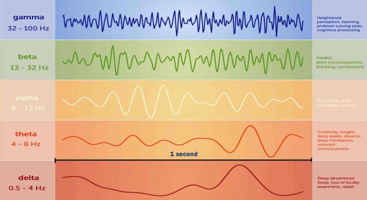
There are five main patterns of brain waves: DELTA, THETA, ALPHA, BETA, and GAMMA. Each brainwave state corresponds to a different state of awareness.
We usually cycle in and out of these different brainwave states throughout the day and night. All brainwave states are essential but must be experienced at the right times and proportions.
Brainwave frequencies are measured in Hertz (Hz), which stands for cycles per second. The greater the Hertz value, the more cycles per second.
When slower brain waves dominate, we can feel sluggish, inattentive, depressed, or dreamy. When higher frequencies dominate, we engage in critical thinking, hyper-alertness, or experience anxiety. However, this can also lead to nightmares, hyper-vigilance, and impulsive behavior.
ROLE AND TYPES OF BRAINWAVES IN NEUROFEEDBACK
Understanding brainwaves, their types, variation patterns, and appropriate clinical manifestations is paramount to organizing effective Neurofeedback training/therapy.
Depending on the types of brainwaves used in neurofeedback training, there are two basic classical directions. One is to focus on low-frequency (alpha or theta) brainwaves in neurofeedback to strengthen relaxation and focus, and the other is to emphasize high-frequency (low beta, beta) brainwaves in neurofeedback to reinforce activation, organize, and inhibit distractibility.
Neurofeedback treatment protocols mainly focus on alpha, beta, delta, theta, and gamma training/treatment or a combination of them, such as the alpha/ theta ratio or the beta/theta ratio.
DELTA BRAINWAVES
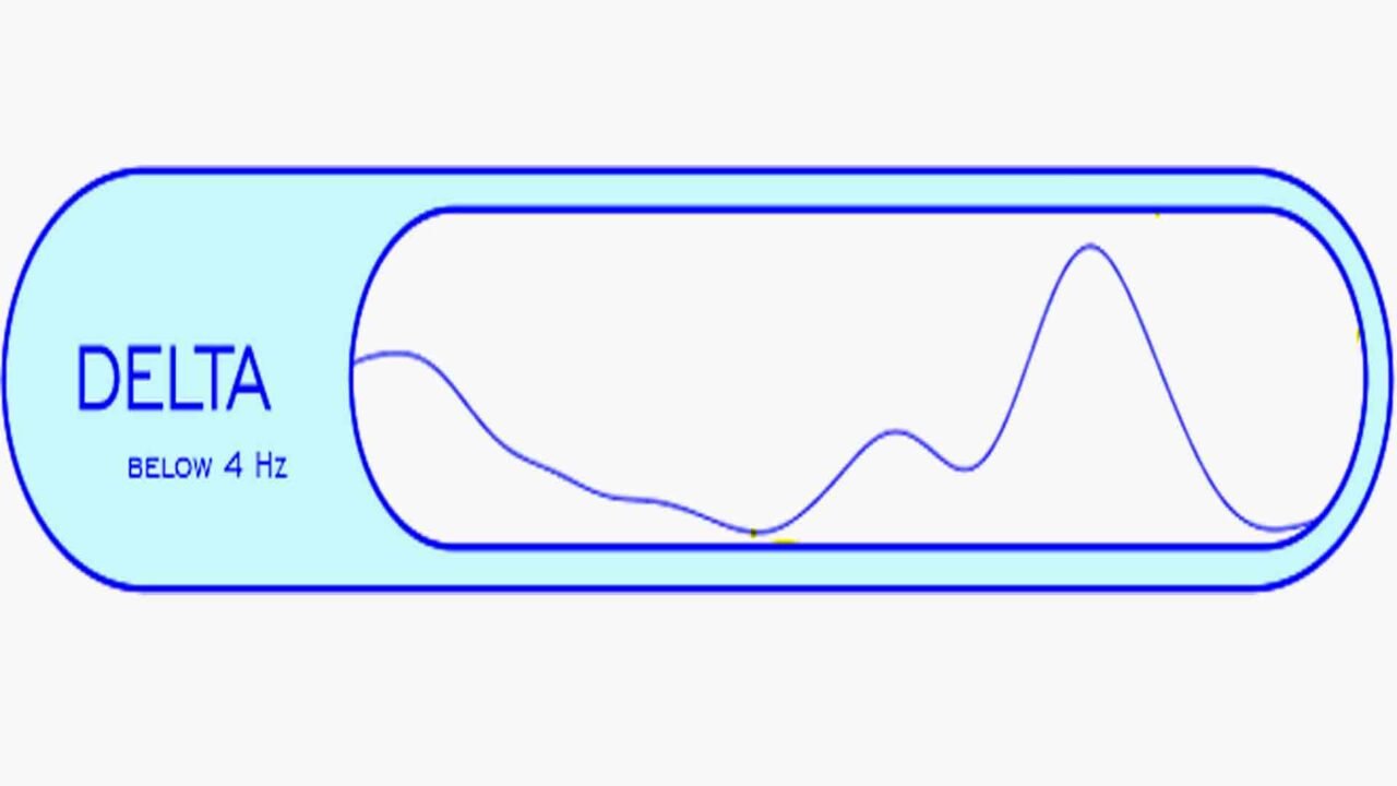
Delta Waves (1-4 Hz) are slow brainwaves that begin to appear in stage 3 of the sleep cycle and, by stage 4, dominate almost all EEG activity. At this stage, healing and regeneration are stimulated and are considered essential for sleep’s therapeutic properties.
An excess of delta waves when a person is awake may result in learning disabilities and Attention Deficit Hyperactivity Disorder (ADHD) and make it extremely difficult to focus. Researchers have found that individuals with different brain injuries often produce delta waves during waking hours, making performing conscious tasks extremely difficult. Sleepwalking and talking typically occur when delta wave production is high. See Neurofeedback protocols here.
THETA BRAINWAVES
Theta waves (4-8 Hz) primarily involve daydreaming and sleep. Cortical theta is commonly seen in young children, but it typically appears in older children and adults during meditative, drowsy, or sleeping states. In theta, we are in a dream with vivid imagery, intuition, and information beyond our everyday conscious awareness. It’s where we hold our fears, troubled history, and nightmares. Theta is our gateway to learning, memory, and intuition. In the theta state, our senses withdraw from the external world and focus on internal signals.
When we are awake, excess theta levels can result in feeling scattered or daydreamy, and this is commonly reported in ADHD. Too much theta in the left hemisphere is thought to result in a lack of organization, whereas too much theta in the right hemisphere results in impulsivity. In individuals with attention disorders, theta waves often appear more prominently towards the front of the brain. See Neurofeedback protocols here.
ALPHA BRAINWAVES
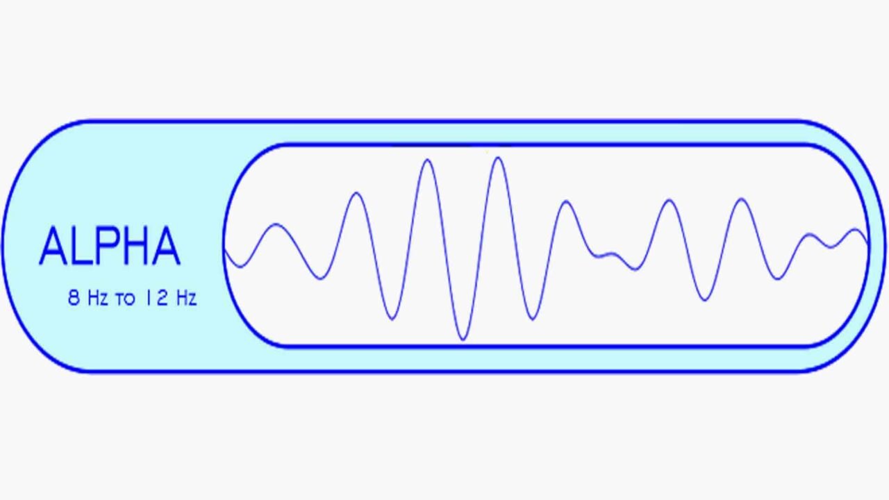
Alpha waves (8-12 Hz) dominate during moments of quiet thought and are similar to meditative states. These brainwaves occur during deep relaxation, typically when the eyes are closed. It is an optimal time to program the mind for success and heightens your imagination, visualization, memory, learning, and concentration. Alpha waves aid mental coordination, calmness and alertness, mind/body integration, and understanding. The brain produces consistently higher magnitudes of alpha waves when the brain is in an idle state. This is why alpha is sometimes referred to as the idle brainwave. Some studies claimed that alpha rhythm has two subsets: lower alpha in the 8-10 Hz range and upper alpha in the 10-12 Hz range. Lower alpha is believed to be related to remembering actions in semantic memory, which is not the case for high alpha.
When the brain is healthy and well-regulated, it produces more alpha waves on the right than on the left. The voice of Alpha is your intuition, which becomes more precise and more profound the closer you get to 7.5Hz.
Alpha rhythm is most prominent in occipital derivations and attenuated by eye-opening. Alpha tends to be highest in the right hemisphere, and too little alpha in the right hemisphere correlates with negative behaviors such as social withdrawal. This phenomenon also occurs in people with depression, especially when they exhibit excessive frontal alpha waves. Alpha waves play a role in actively and adequately inhibiting irrelevant sensory pathways. See Neurofeedback protocol here.
BETA BRAINWAVES
Beta waves (12-30 Hz) represent the normal waking state of consciousness, mainly when attention focuses on cognitive tasks and the outside world. Additionally, beta waves feature fast-wave activity and dominate during moments of alertness, attentiveness, problem-solving, decision-making, and other focused mental tasks.
Low beta, or so-called Sensorimotor Rhythm (SMR) (12-15 Hz), is thought to be ‘fast idle’ or ‘musing thought. ‘ It reflects mental alertness and physical relaxation and is closely related to readiness for action and attentive listening.
Beta (15-20 Hz) reflects high engagement, actively figuring things out, thinking, focusing, sustained attention, tension, alertness, and excitement.
High Beta (20-30 Hz) is a highly complex thought that integrates new experiences, high anxiety, or excitement. Continuous high-frequency processing is not an efficient way to run our brains, and it can result in tension and difficulties in relaxing. If present at night, it can also result in problems settling the mind and falling asleep.
Beta waves dominate the left hemisphere, while excessive beta activity in the right hemisphere can correlate with mania.
The brain consistently generates higher levels of beta waves when it focuses externally, remains alert, and engages in critical reasoning, thought, and concentration. Beta waves represent the fastest processing speed, and when the brain functions well, it produces more beta waves on the left than on the right.
While Beta brain waves are essential for effective functioning throughout the day, they can also translate into stress, anxiety, and restlessness. See the Neurofeedback protocol here.
GAMMA BRAINWAVES
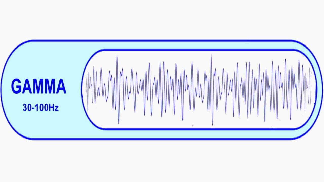
This brainwaves (30-100Hz) have the highest frequencies of any brainwave, oscillating between 30 to 100 Hz.
Gamma waves are associated with peak concentration and high levels of cognitive functioning. Researchers observe a synchronous burst of gamma bandwidth during “ah-ha” moments.
Low levels of gamma activity are associated with learning difficulties, impaired mental processing, and limited memory. In contrast, high gamma activity correlates with a high IQ, compassion, excellent memory, and happiness.
Gamma brainwaves hold limited clinical value because researchers argue that existing EEG technology cannot effectively measure them due to muscle contamination. Although promising studies indicate that gamma training can enhance intelligence, it won’t be used correctly in clinical settings until researchers resolve this technology issue.
ELECTROENCEPHALOGRAPHY. ELECTRODE PLACEMENT SYSTEM. REGISTRATION OF BRAINWAVES IN NEUROFEEDBACK
An electroencephalogram (EEG) represents the brain’s electrical activity. The EEG signals have deficient voltage amplitude levels ranging from 1 to 100 microvolts. They also have very low frequencies, ranging from 1 Hz to 100 Hz, as described above. ECG equipment amplifies the potentials received from the sensors hundreds of thousands of times and writes them into the computer’s memory. The EEG waveforms differ in shape and show dramatic changes in the individual’s state.
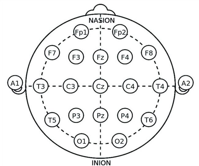
The location of the electrodes is essential when registering EEG, and the electrical activity simultaneously recorded from different points of the head can vary greatly. The most popular scheme used in the placement of electrodes for the EEG picks is the international 10-20 electrode placement system. As the cranial area is divided into four main regions, the electrodes are placed accordingly in the different areas.
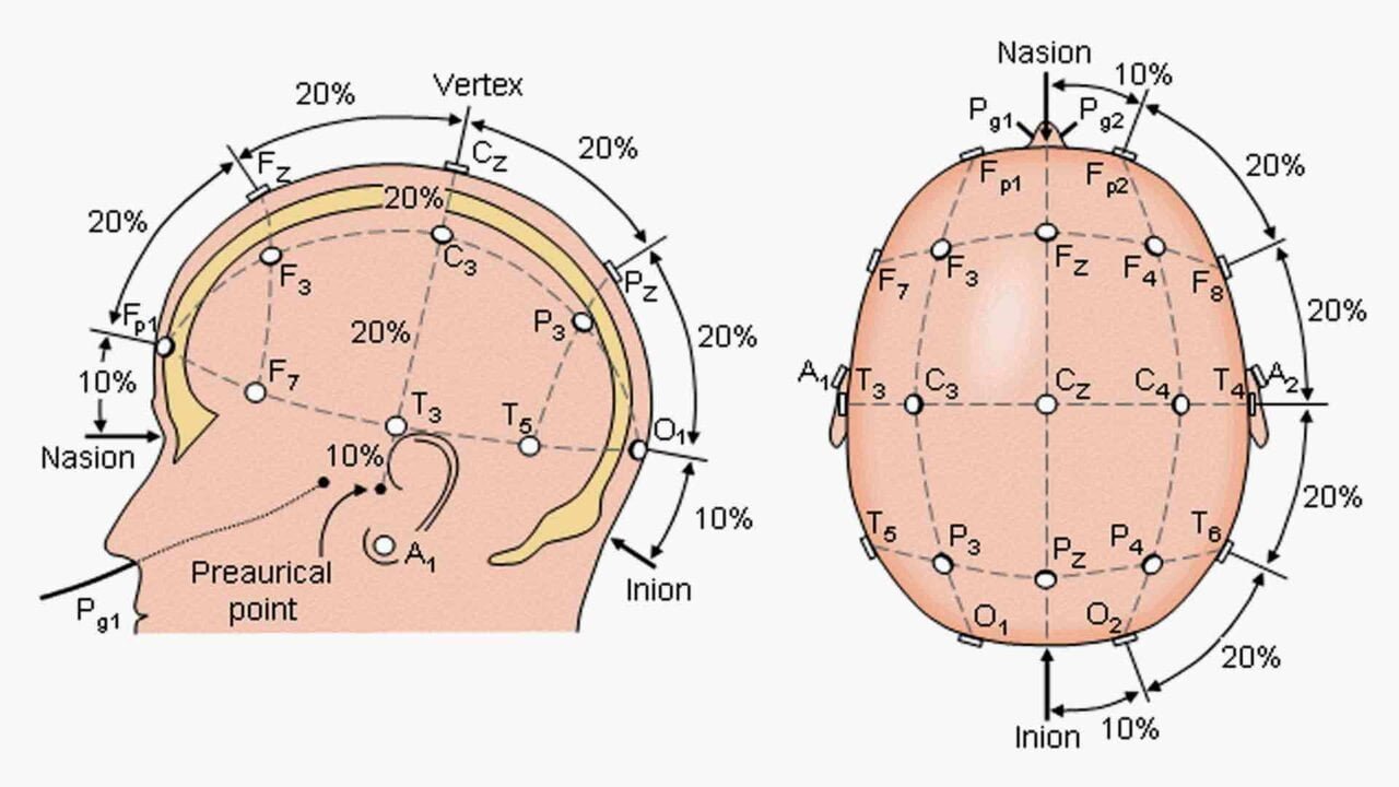
The main regions of the skull are frontal, parietal, temporal, and occipital regions. Electrodes placed in the different areas are labeled as F (Frontal), P (Parietal), T (Temporal), and O (Occipital). Odd numbers correspond to the left side of the brain, while even numbers indicate the right side. In this setup, the head is mapped using four standard points: the nasion, inion, and left and right ears. The letter “z,” as in Pz, indicates that the scalp location lies along the central line between the nasion and inion. Additionally, Fp1 and Fp2 refer to the left and right poles of the forehead, respectively.
10-20 electrode placement system
In the 10-20 electrode placement system, we use 20 electrodes. One electrode is used to ground the subject. When recording EEG, there are two main methods of electrode application: bipolar and unipolar.
In a unipolar arrangement, one electrode sits at a point traditionally considered electrically neutral, such as the left or right earlobes (A1/A2) or the nose bridge. This electrode acts as the standard reference for all channels. In a unipolar recording, the ECG reflects the activity of one of the points/leads relative to electrically neutral points/leads (for example, the activity of the frontal or occipital leads relative to the earlobe). So, to measure the bioelectric potential from the brain, we use any of the nineteen electrodes and the common electrode.
In a bipolar system, both electrodes are placed in electrically active scalp points, and any of the two electrodes is used for the measurement. In the bipolar recording, an EEG record reflects the result of the interaction of two electrically active points (for example, frontal and occipital leads).
The choice of one or another recording variant depends on the study’s objectives. In research practice, the unipolar registration option enjoys wider use because it allows researchers to study the isolated contribution of a specific brain area to the learning process.
Before placing the electrodes, researchers measure the nasion-inion distance. Next, they mark points on the head at 10%, 20%, 20%, 20%, 20%, and 10% of this length. They then place the electrodes on these marked points. This system is known as the 10-20 electrode system.
BRAINWAVES IN NEUROFEEDBACK. MEDICAL AND NON-MEDICAL APPLICATION OF NEUROFEEDBACK
Understanding Brainwave Regulation
The brain typically uses environmental cues to regulate its brainwave patterns. However, when certain patterns become overly dominant or dysfunctional, they can lead to brainwave dysregulation and associated behavioral problems. Researchers identify brainwave dysregulation by detecting abnormal brainwave ratios and measuring them with EEG equipment. In this context, neurofeedback emerges as a valuable form of biofeedback, enabling individuals to adjust deregulated brainwave rhythms actively.
The Applications of Neurofeedback
EEG biofeedback teaches individuals to control their brainwaves to achieve a desired state consciously. Neurofeedback serves both medical and non-medical purposes, although the distinction can be subtle. Non-medical applications primarily focus on personal improvement and brain conditioning, enhancing relaxation, attention, focus, concentration, and self-awareness. Furthermore, individuals often use it alongside meditation, counseling, or hypnosis to reach altered states of consciousness, frequently without professional assistance. However, anyone seeking to address medical symptoms should consult a healthcare professional. Detailed information about the indications for neurofeedback use and methods for peak performance is available on our website.
The Role of Neuroplasticity
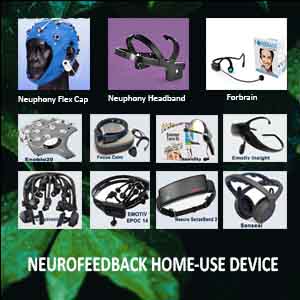
Neurofeedback home-use systems are designed for recreational, educational, or entertainment purposes and are not classified as medical instruments. Our site provides comprehensive information about various neurofeedback devices for home use. Nevertheless, a device becomes medical if it claims to offer direct benefits for relaxation or symptom relief from disorders.
Importantly, although neurofeedback can enhance attention and concentration, it might be perceived as a medical procedure for those with suspected or diagnosed Attention Deficit Disorder (ADD). In such cases, one could argue that neurofeedback aimed at alleviating ADD symptoms constitutes a medical intervention, particularly when reducing reliance on stimulants like Ritalin is desired.
Conversely, when parents, teachers, or counselors use neurofeedback in a home or educational setting, their goal shifts to helping the child achieve relaxed attentiveness. In this context, the practice may be considered educational rather than therapeutic. Ultimately, neurofeedback leverages the brain’s capacity for change through neuroplasticity. It mirrors the learning process of acquiring new skills by forming connections between nerve cells and enhancing pathways within the brain. As individuals utilize these pathways more frequently, their performance improves.
Essentially, neurofeedback provides the ideal learning conditions by promoting awareness of healthier brainwave patterns and reinforcing positive changes throughout training sessions. For more detailed information about the types of neurofeedback training available, please explore our resources.

