BRAIN REGION FUNCTION IN NEUROFEEDBACK
Neurofeedback is a powerful tool that leverages the brain’s self-regulating ability by targeting specific brain regions. Understanding brain region function in neurofeedback is crucial for optimizing its effectiveness. Each brain region uniquely controls thoughts, emotions, and behaviors, making it essential to focus on the right areas during training. By tapping into this knowledge, neurofeedback practitioners can enhance cognitive performance, emotional stability, and mental clarity. This article explores the key brain regions involved in neurofeedback, shedding light on their specific functions and how they contribute to improved brain health.
Table of Contents
ToggleThe Structure and Functions of the Human Brain
The human brain is not only one of the most essential organs in the human body; it is also the most complex. The brain is the central part of the nervous system, which governs the functions of various organs in the body.
The brain is composed of the cerebrum, cerebellum, and brainstem.
The cerebrum is the most significant part of the brain. It contains the cerebral cortex of the two cerebral hemispheres and several subcortical structures, including the hippocampus, basal ganglia, and olfactory bulb. The cerebrum performs higher functions like interpreting touch, vision, hearing, speech, reasoning, emotions, learning, and fine control of movements.
The cerebellum is located under the cerebrum. Its function is to coordinate muscle movements and maintain posture and balance.
The brainstem is a relay center connecting the cerebrum and cerebellum to the spinal cord. It performs many automatic functions, such as breathing, heart rate, body temperature, wake and sleep cycles, digestion, sneezing, coughing, vomiting, and swallowing.


Hemispheres of the Brain and the Cerebral Cortex
The cerebrum consists of two halves: the right and left hemispheres. A bundle of fibers joins them called the corpus callosum, which transmits messages from one side to another. Each hemisphere controls the opposite side of the body.
The left hemisphere (LH) is usually the dominant hemisphere. It is responsible for activities on the right side of the body. The LH is good at logic and analytical reasoning. Two main language centers are in the LH: Broca’s area, which deals with verbal expression and speech production, and Wernicke’s area, which deals with verbal comprehension. The left hippocampus stores verbal memories. Only around 20% of left-handed people are right-hemisphere dominant.
The right hemisphere (RH) is the non-dominant hemisphere. It is responsible for activities on the left side of the body. The Right Hemisphere is involved in creativity, perception, visual-spatial processing, and facial recognition.
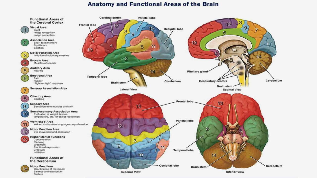
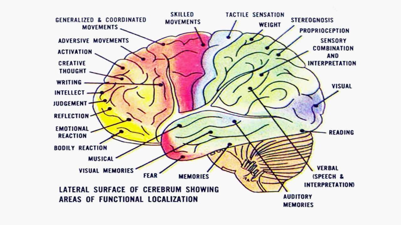
The cerebral cortex is the outer layer of neural tissue (grey matter) covering the surface of the cerebrum. The cerebral cortex is the part of the brain that makes humans unique. Distinctly human traits, including higher thought, language, and human consciousness, as well as the ability to think, reason, and imagine, all originate in the cerebral cortex. The cerebral cortex is what we see when we look at the brain. The outermost part of the brain is divided into four lobes: the frontal, parietal, occipital, and temporal lobes. Each lobe handles distinct functions, such as reasoning and auditory perception.

FUNCTIONAL REGIONS AND AREAS OF THE CEREBRAL CORTEX IN NEUROFEEDBACK
Functional Areas of the Cerebral Cortex
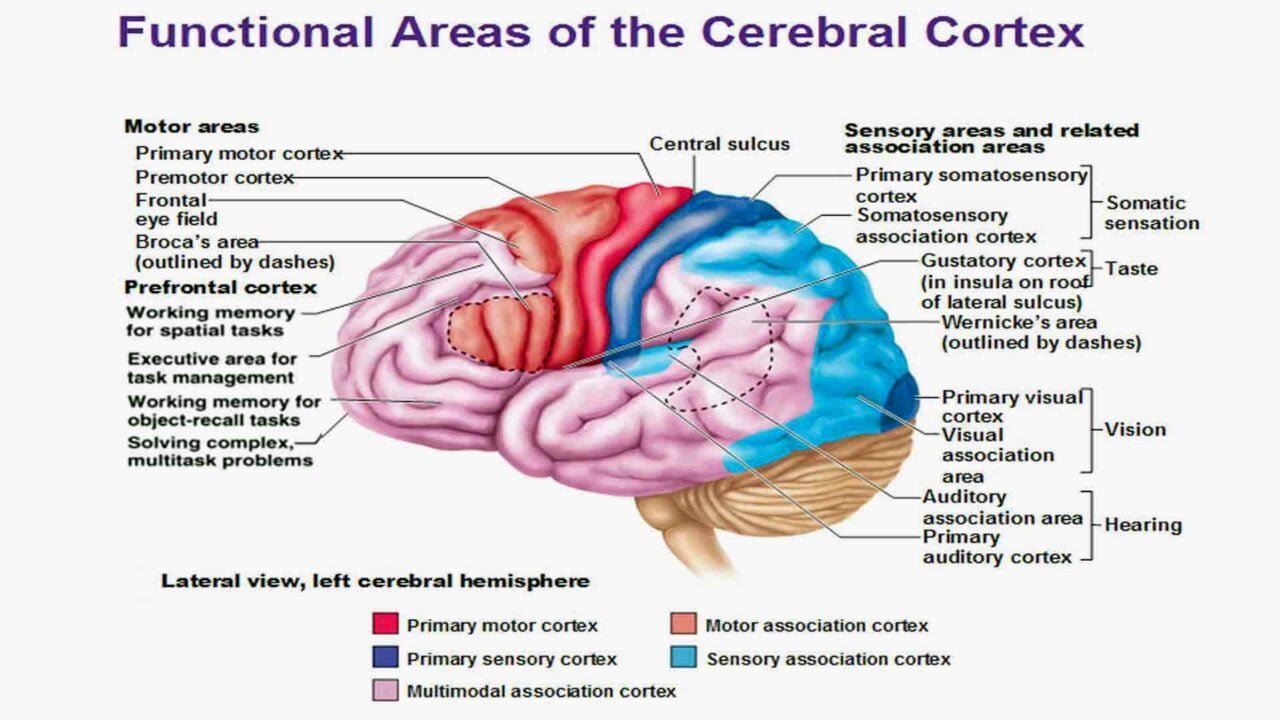
The cerebral cortex has two functional areas: sensory and motor areas. The precentral gyrus is entirely motor, while the postcentral gyrus is entirely sensory.
The three types of functional areas are:
Motor areas – allow you to act upon a sensation
- Premotor Cortex – plans movements;
- Primary Motor Cortex – sends signals to generate movements;
- Two special motor cortices (Frontal Eye Field, Broca’s area)
Sensory areas
- Primary Sensory Cortex – makes you aware of a sensation;
- Association areas – give meaning to/make associations with a sensation;
- Multimodal Association Areas – make associations between different types of stimuli.
The Role of Association Areas in Sensory Integration
Association areas in the cerebral cortex integrate and interpret sensory information. Each sensory modality—sight, sound, or touch—has a primary sensory cortex corresponding to the association area. These areas link new sensations with past experiences to give them meaning.
Multimodal Association Areas
These large areas of the cerebral cortex receive sensory input from multiple sensory modalities and various association areas and help make associations between several types of sensory info. We have three multimodal association areas: Posterior, Anterior, and Limbic association areas.
The visual, auditory, and somatosensory association areas meet in the posterior association area, which gives us spatial awareness of our body. For example, the kinesthetic sense is powerful in professional dancers or athletes. Wernicke’s area on the left side deals with reading and naming things. On the right side, this area helps us understand emotional overtones/undertones of speech (the multiple ways of saying “I’m fine”).
The anterior association area includes the prefrontal cortex. This part of the brain receives information from the posterior association area and helps integrate that information with experience with the help of the limbic association area. It has a lot to do with thinking and making judgments, and it’s where you understand what’s socially acceptable, how to behave, and anything related to heavy human conditioning.
The limbic association area is located on the medial side of the frontal lobe. It helps form memories, translate those to motor responses, process emotions, and guide emotional responses. It’s crucial for social interactions and expressions of the personality.
The role of brain region function in neurofeedback training organization is paramount and essential for effective therapy.
FRONTAL LOBE BRAIN REGION FUNCTION IN NEUROFEEDBACK
The Frontal Lobe: Functions and Importance
The frontal lobe, located at the front of the brain, is essential for various cognitive and motor functions. It governs immediate and sustained attention, time management, working memory, executive planning, and initiative. In addition to reasoning, motor skills, and higher-level cognition, the frontal lobe plays a crucial role in expressive language.
Primary Motor Cortex and Movement
The primary motor cortex lies at the back of the frontal lobe, anteriorly to the central sulcus. This area of the brain receives information from various lobes and utilizes this information to carry out body movements. It works in association with pre-motor regions to plan and execute movements. The frontal lobes are heavily connected to the amygdala, which deals with emotions. The frontal lobes and amygdala work together as part of the social brain’s network, dealing with empathy and social skills.
- The lesion in this area will result in paralysis/paresis (partial loss of movement or impaired movement) of the contralateral body area.
Premotor Cortex: Planning Complex Movements
The premotor cortex is anterior to the primary motor cortex and receives processed sensory information. This helps to plan complex movements and sends the plan to the primary motor cortex. It is responsible for sensory guidance of movement and control of proximal and trunk muscles of the body.
- The lesion will result in Apraxia (loss of the ability to execute or carry out learned purposeful movements despite having the desire and the physical ability to perform the movements).
Frontal Eye Field: Controlling Eye Movements
The frontal eye field is anterior to the premotor cortex and helps control voluntary eye movements. Stimulation of the frontal eye field on one side produces conjugate eye movement on the contralateral side.
- Lesion produces a transient deviation of eyes to the ipsilateral side and paralysis of the contralateral gaze.
Supplementary Motor Area: Coordinating Complex Movements
Stimulation of the supplementary motor area produces posturing responses such as turning the head and eyes toward the moving arm and programming for complex movements involving several body parts.
Broca’s Area: Language and Speech Production
Broca’s area is located only in the left cerebral hemisphere. It is anterior to the premotor cortex, near the auditory sensation areas. It is what helps control motor movements for speech production. The corresponding area on the right side controls the emotional overtones of spoken words. In other words, it helps dictate how you say/form the words in your speech. It is responsible for language production and comprehension (It directly connects with the muscles responsible for the movements of the tongue, lips, vocal cord, and pharynx).
- The lesion will cause motor aphasia (a language disorder in which there is an impairment, but not loss of speech and comprehension of speech) but only when the dominant hemisphere is involved. The patient knows what he wants to say, but speech is slow, deleting many prepositions and nouns, certain words are skipped and missed, or words are repeated.
Prefrontal Cortex: Cognitive and Emotional Regulation
The prefrontal cortex is located in the anterior portion of the frontal lobe, takes nearly 1/4 of the cortex, and performs cognitive functions. Regulating the flow of sensory information plays a crucial role in the posterior sensory systems. Originating in the basal ganglia and motor cortical/subcortical areas, it is responsible for planning, initiating, and inhibiting actions. The prefrontal cortex involves intellect, cognition, planning, judgment, problem-solving, conceptualizing, recall, and personality. It is necessary for judgment, reasoning, persistence, and conscience. It is also related to mood and closely linked to the limbic system (the emotional part of the brain).
- Lesion Impact: Damage to the Prefrontal Cortex can result in a loss of initiative, changes in social behavior, and altered decision-making abilities. It may also cause people to dress carelessly or exhibit socially inappropriate behavior.
Damage to the frontal lobe can lead to changes in sexual habits, socialization, and attention, as well as increased risk-taking. These changes underline the frontal lobe’s pivotal role in regulating both cognitive and emotional aspects of behavior.
FRONTAL LOBE NEUROFEEDBACK APPLICATION
Neurofeedback to Fz and Fpz can impact social behavior. Weaknesses in this area are associated with oppositional defiance disorder and antisocial behaviors. Slowing may be evident in these frontal areas. Neurofeedback training in the right prefrontal cortex may lead to reduced fear and a sense of calm and well‐being due to the connections with the amygdala. Frontal issues tend to present as patients being in a fog, struggling to focus, getting into trouble, having social issues, and being fearful, unmotivated, and disconnected.
Left hemisphere: neurofeedback training application in Fp1, F3, and F7 improves working memory and concentration and increases executive planning performance and positive emotions.
Right hemisphere: neurofeedback application in Fp2, F4, and F8 improves episodic memory and social awareness.
PARIETAL LOBE BRAIN REGION FUNCTION IN NEUROFEEDBACK
The Parietal Lobe: Sensory Processing and Problem Solving
The parietal lobe is located in the middle section of the brain and is associated with processing tactile sensory information such as pressure, touch, and pain. A portion of the brain known as the somatosensory cortex is located in this lobe and is essential to processing the body’s senses.
Functional Differences Between the Left and Right Parietal Lobes
The parietal lobes solve problems that the frontal lobes conceptualize. The left parietal lobe deals with complex grammar, object naming, sentence construction, and aspects of math. The right parietal lobe deals with the more spatial aspects of math. The right parietal lobe also deals with map orientation, right-left recognition, and spatial recognition.
Primary Somatosensory Cortex: Conscious Sensory Awareness
Located in the postcentral gyrus, the Primary Somatosensory Cortex plays a key role in the conscious awareness of general somatic senses, such as touch and pressure. It receives sensory information from the skin and skeletal muscles, allowing for precise spatial discrimination. This region processes input from the opposite side of the body, known as contralateral projection.
- Stimulation Impact: Stimulation of the Primary Somatosensory Cortex produces contralateral tingling or numbness but does not cause pain.
- Lesion Impact: Damage to this area can result in a contralateral loss of tactile discrimination and position sense, but it does not relieve pain.
Somatosensory Association Cortex: Integrating Sensory Input
It is located posterior to the primary somatosensory cortex. It integrates sensory information to form a comprehensive understanding of the stimulus and determines size, texture, and the relationship of parts.
- Primary Gustatory Cortex: Responsible for the perception of taste.
- Lesion Impact: Damage to this region can lead to contralateral (mostly) ageusia (loss of taste perception).
Parietal Association Cortex: Higher-Level Processing
The Parietal Association Cortex processes more complex sensory information, helping integrate data from various senses.
- Parietal Neglect Syndrome: Damage to the parietal lobe may result in parietal neglect syndrome, where a person fails to recognize the side of the body opposite the injury. For instance, they may not bathe, shave the affected side, or even deny their limbs’ existence. Individuals might ignore objects in the contralateral visual field.
- Example: A lesion on the left hemisphere would cause neglect on the right side, where a person might forget to shave the right side of their face or ignore it entirely.
Impact of Parietal Lobe Damage
Damage to the parietal lobes can make it difficult to attend to multiple objects simultaneously across both sides of the visual field. It may also impair the ability to perceive spatial relationships and interfere with recognizing familiar objects through touch (astereognosis).
PARIETAL LOBE NEUROFEEDBACK APPLICATION
Left hemisphere: application of neurofeedback training in P3 gives the possibility to regulate problem-solving, increase mathematical and complex grammar performance, and increase attention.
Right hemisphere: neurofeedback training in P4 improves Spatial awareness and geometry task performance.
Training the left and right parietal lobes, P3 and P4, is good for solving the dyscalculia problem.
TEMPORAL LOBE BRAIN REGION FUNCTION IN NEUROFEEDBACK
The Temporal Lobe: Auditory Processing and Memory
It is located on the lateral bottom section of the brain. This lobe also contains the primary auditory cortex, vital for interpreting sounds and the language we hear. The left temporal lobe handles verbal memory, while the right temporal lobe focuses on music. The temporal lobe includes the auditory cortex (involved in processing auditory information) and the hippocampus (involved in memory). The left temporal lobe is responsible for word recognition, whereas the right temporal lobe handles facial recognition.
Primary Auditory Cortex: Sound Processing
It sits in the upper part of the temporal lobe and processes auditory information such as pitch, rhythm, and loudness.
- The lesion makes it challenging to recognize the distance and direction of sound, especially when sound comes from the contralateral side.
The Auditory Association Cortex: Understanding Sounds
Located just behind the primary auditory cortex, the Auditory Association Cortex stores memories of sounds and permits their perception. It is also responsible for language understanding, recognition, and formulation and is closely linked to Wernicke’s area.
- Damage can result in aphasia (the inability to express oneself in terms of speech). This is receptive aphasia; the person will be unable to comprehend spoken words and speak fluently but without any meaning.
Limbic Temporal Cortex: Emotions and Memory
Three essential structures reside on the medial surface of the temporal lobe: the olfactory cortex, amygdala, and hippocampus. These structures control visceral function, emotions, behavior, and memory.
- Stimulation Impact: Stimulating the Limbic Temporal Cortex can elicit memories of past events.
- The left posterior area stores verbal information and memories.
- The right posterior area stores visual information and memories.
- Lesion Impact: A bilateral lesion of the limbic temporal cortex causes prosopagnosia (loss of facial recognition). Alzheimer’s disease commonly affects this area, and a lesion here can erase all memories.
Primary Olfactory Cortex: The Smell Center
On top of the cribriform is the nasal foramina, which hit the olfactory bulb and then ran toward the primary olfactory cortex through the olfactory tract. This cortex is where you get a sensation of smell before you’ve figured out what the scent is. The olfactory cortex is located on the medial aspect of the temporal lobe, in the uncus (aka piriform lobe). The olfactory cortex is also called the Rhinencephalon or “nose brain.” This is the most primitive part of the cerebrum and connects directly to the limbic system (emotional system), which is why smells often directly trigger emotions and our most profound memories.
Amygdala: Emotional Memory
The amygdala is responsible for emotional responses and memory, primarily fear-related. It also processes mood and conscious emotional reactions to positive or negative events.
Hippocampus: Memory Formation
The hippocampus forms declarative memories (such as facts and events). However, it is not involved in working memory, procedural memory, or memory storage. Damage to the hippocampus impairs the formation of new declarative memories but does not affect existing memories.
Impact of Temporal Lobe Damage
Damage to the temporal lobe can lead to problems with
- Memory formation
- Speech perception
- Language skills.
TEMPORAL LOBE NEUROFEEDBACK APPLICATION
OCCIPITAL LOBE BRAIN REGION FUNCTION IN NEUROFEEDBACK
The occipital lobe is located at the back portion of the brain and is associated with interpreting visual stimuli and information. The primary visual cortex, which receives and analyzes data from the eyes’ retinas, is in the occipital lobe.
The primary visual cortex is located on the posterior part of the occipital lobe and receives visual information from the retinas. It provides macular vision (vision in which each eye is used separately). Compared to binocular vision, macular vision increases the field of view while limiting depth perception.
- The lesion causes homonymous hemianopsia (partial blindness resulting in a loss of vision in the same visual field of both eyes).
The visual association area surrounds the primary visual cortex and interprets visual stimuli (e.g., color, form, and movement).
Parastriate cortex The Peristriate cortex receives visual info bilaterally and complex processing for color, movement, direction, and visual interpretation.
- The lesion can cause visual agnosia (a loss of ability to recognize objects, persons, sounds, shapes, or smells), while the specific sense is not defective, and there is no significant memory loss.
Damage to the occipital lobe can cause visual problems such as difficulty recognizing objects, an inability to identify colors, and trouble remembering words.
Traumatic memories that accompany visual flashbacks are processed in the occipital lobes.
OCCIPITAL LOBE NEUROFEEDBACK APPLICATION
Neurofeedback training in O2 helps maintain performance in areas such as counting and auditory discrimination, increasing the quality of musical performance.
Depression and anxiety have also been shown to remit after deep‐state neurofeedback training in Oz and O1.
Visual memories, accurate reading, and traumatic memories accompanying visual flashbacks are usually processed in the occipital lobes, O1, O2, and Oz.
Patients suffering from PTSD (Post Traumatic Stress Disorder) may benefit from neurofeedback training in the occipital lobes, mainly when doing deep-state training.
BRAIN REGIONS FUNCTION IN NEUROFEEDBACK
Neurofeedback: Rewiring the Brain for Optimal Functioning
Certain brain parts can quickly get out of whack through specific life experiences. This causes an over or underproduction of brain waves in particular brain regions, which leads to negative symptoms. For example, a trauma survivor would have overactivation in the amygdala, which causes him/her to constantly be on high alert, overly vigilant, and fearful. In addition, a trauma survivor would have under-activated prefrontal cortexes and anterior cingulate cortexes.
Neurofeedback helps rewire the brain, calming the overactivated regions and bringing online the under-activated areas, allowing the brain to function and operate as it should.
Electrodes are attached to the client’s head in the appropriate brain regions that need training. Once connected, the information and brainwaves are displayed on a screen in real time, giving the brain a mirror to understand its function better.
How Neurofeedback Works
Neurofeedback will help direct the brain to create more of some frequencies and less of others. This process creates new patterns and pathways in the brain, leading to healthier reactions to stress and self-regulation. As these new frequencies and pathways develop, the brain is rewarded through the neurofeedback program, incentivizing the brain to manifest these pathways more frequently. Once the brain has relaxed, it can make better choices on how to react to stress. It’s no longer hijacked by fear and stress, allowing access to the prefrontal cortex to make more logical, thought-out decisions.
Providing individuals with real-time feedback about the level of brain activity or rhythm in different regions of brain neurofeedback can potentially help them learn to control the activation of specific brain regions. Specifically, the training can be customized for patients depending on the type of pathology involved; the feedback can be explicitly programmed to up or down-regulate the activity in a specific brain region as appropriate.
Specific Benefits of Neurofeedback by Brain Region
Neurofeedback provided in the frontal lobe can impact social behavior. Weaknesses in this area are associated with oppositional defiance disorder and antisocial behaviors. Slowing may be evident in these frontal areas.
Neurofeedback training in the right prefrontal cortex may reduce fear as well as a sense of calm and well-being due to the connections with the amygdala. Frontal issues tend to present as patients being in a fog, struggling to focus, getting into trouble, having social issues, fearful, unmotivated, and disconnected.
Neurofeedback in the left parietal lobe can help with problem-solving, math, complex grammar, and attention improvement. In contrast, neurofeedback training in the right parietal lobe can improve spatial awareness and geometry skills. For dyscalculia, it is helpful to train both the left and right parietal lobes.
Neurofeedback training in the left temporal region can improve word recognition, reading abilities, and language skills. It can also enhance memory and train for dyslexia. Meanwhile, neurofeedback training in the right temporal region improves object recognition, music skills, social cues, and facial recognition.
A Lasting Solution
The best part about neurofeedback is that no further treatment is needed when the brain has created new, positive pathways. Rather than influencing the chemical reactions in the brain through pharmaceutical drugs, neurofeedback changes the fundamental brain activity. Instead of treating a symptom through daily medication, it’s an opportunity to treat the underlying issue within the brain.
Guidance through Brain Mapping (qEEG)
Since different brain regions contribute to various symptoms, a brain map or quantitative electroencephalogram (qEEG) is often used to guide neurofeedback treatment. This map helps identify which areas require attention, ensuring a more targeted and effective approach. However, it’s important to note that no treatment is universally effective, and like all therapies, neurofeedback may have potential risks. We recommend discussing this treatment with a knowledgeable professional to make an informed decision for yourself or your child.
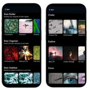
Applications of Neurofeedback
Neurofeedback is used for both medical and non-medical purposes. Non-medical applications focus on personal improvement and brain conditioning—enhancing relaxation, attention, focus, concentration, and self-awareness. It can also be used as an adjunct to practices like meditation, counseling, or hypnosis, and it can aid in achieving altered states of consciousness. Neurofeedback can even be practiced without professional intervention for these purposes.
Our website provides detailed information on the indications for neurofeedback use, methods for achieving peak performance, and its effectiveness in treating various disorders. You’ll also find comprehensive descriptions of different neurofeedback devices for home use.
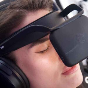
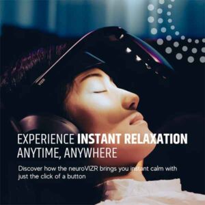
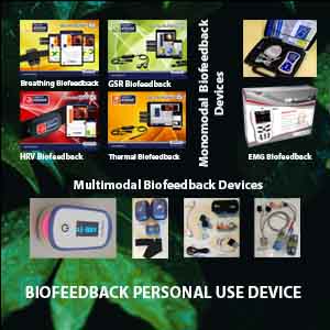
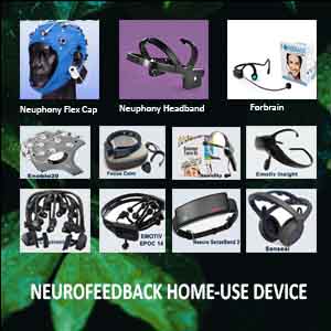
NEUROFEEDBACK HOME USE DEVICE
Medical and Non-Medical Uses of Neurofeedback Devices
Neurofeedback devices are used for both medical and non-medical purposes, and the distinction between the two can sometimes be subtle. In non-medical applications, neurofeedback primarily focuses on personal improvement and brain conditioning. It aims to enhance relaxation, attention, focus, concentration, and self-awareness. It can also serve as an adjunct to practices such as meditation, counseling, hypnosis, or achieving altered states of consciousness. In these cases, neurofeedback can often be used without professional intervention.
However, professional guidance should be sought when neurofeedback addresses medical conditions, such as relieving symptoms associated with a disorder.
Non-Medical vs. Medical Devices
Neurofeedback systems designed for recreational, educational, or entertainment purposes are not classified as medical instruments. These devices allow the user to control a computer or engage in other activities for fun or learning. Our site provides detailed information regarding home-use neurofeedback devices and their non-medical applications.
However, devices marketed to provide direct benefits for relaxation or relieve symptoms of disorders are considered medical devices.
Many of the same functions and capabilities are present in non-clinical settings, but they are presented within the context of educational or recreational use. Despite the differences in labeling and claims, the actual benefits for the user can be similar in both medical and non-medical contexts, depending on how the device is applied.
Key Difference Between Medical and Non-Medical Devices
The primary difference between medical and non-medical embodiments of neurofeedback devices lies in the claims and the user’s intent and expectations.
For instance, neurofeedback can improve attention and concentration, which could be seen as a personal improvement application. However, if a person with Attention Deficit Hyperactivity Disorder (ADHD) uses neurofeedback to relieve symptoms, the procedure would be classified as medical.
When neurofeedback is explicitly used to reduce ADHD symptoms or assist with reducing dependency on stimulants like Ritalin, it is considered a medical treatment. On the other hand, if a parent, teacher, or counselor uses neurofeedback in a home or educational setting to help a child achieve relaxed attentiveness and improve academic performance, it would be regarded as education, not treatment.
Neuroplasticity and Neurofeedback
Neurofeedback leverages the brain’s ability to change and adapt through a process known as Neuroplasticity. This is the same process that occurs when we learn new skills. The brain learns and improves performance by connecting nerve cells and utilizing critical pathways linking different brain regions.
The more often these pathways are activated, the more efficiently the brain performs associated tasks.
Neurofeedback operates on the principle of operant conditioning, where behaviors are shaped by their consequences. Neurofeedback provides ideal learning conditions by:
- Facilitating awareness when the brain produces healthier brainwave patterns.
- Offering reinforcement for positive changes.
- Allowing multiple opportunities for practice within a training session.
Through regular neurofeedback sessions, the brain learns to create and reinforce positive neural patterns, resulting in lasting improvements in brain function.