HRV Training

HRV Training. How to improve performance

In recent years, integrating HRV training (Heart Rate Variability) with biofeedback guidance has emerged as a promising avenue for enhancing performance across various domains. This approach offers individuals real-time insights and cues to optimize their HRV metrics and overall performance outcomes.

HRV, the variation in the time interval between heartbeats, is a window into the autonomic nervous system’s activity, reflecting the balance between sympathetic and parasympathetic influences. Individuals can use targeted interventions, such as controlled breathing exercises and mindfulness techniques, to modulate their HRV. This modulation can then influence their physiological and psychological states.

Introduction to HRV Training

Heart Rate Variability (HRV) training aims to improve the function of the autonomic nervous system. It works by modulating the variation in the time intervals between heartbeats. This variability indicates the balance between sympathetic (fight-or-flight) and parasympathetic (rest-and-digest) nervous system activity. HRV training typically involves controlled breathing exercises, meditation, and relaxation techniques influencing HRV patterns. By enhancing HRV, individuals can potentially improve their resilience to stress. They may also experience better recovery and optimize performance in various aspects of life.

HRV Indexes

A high HRV index indicates more significant variability between heartbeats. This suggests a well-regulated and flexible autonomic nervous system. Such regulation is associated with better health outcomes and greater resilience to stress. A high HRV index typically falls within the 50-100 milliseconds range. However, specific values can vary based on age and overall health status.

A low HRV index reflects reduced variability between heartbeats, indicating a more rigid and less adaptable autonomic nervous system. Lower HRV often correlates with an increased risk of health issues, including cardiovascular disease, anxiety disorders, and poor emotional regulation. Typically, a low HRV index is below 50 milliseconds. Monitoring HRV indices offers valuable insights into an individual’s physiological and psychological well-being. Understanding these indices can guide interventions aimed at improving overall health and performance.

HRV training leverages the body’s ability to adapt and respond to stressors. Consistent practice allows individuals to train their autonomic nervous system to achieve better balance and coherence. This results in increased variability in heart rate patterns. Consistent practice can improve cardiovascular health, emotional regulation, cognitive function, and well-being.

Indications for using HRV training

Experts may recommend HRV training as a complementary therapy for conditions such as hypertension, anxiety disorders, depression, and chronic pain. These conditions often involve dysregulation of the autonomic nervous system.

Stress Management:

HRV training helps individuals cope with stress by promoting relaxation and reducing its physiological effects on the body. Improved HRV links to greater resilience to stressors and better emotional regulation.

Athletic Performance Enhancement:

HRV training can benefit athletes by optimizing performance and enhancing recovery. It also helps manage the demands of training and competition more effectively. Enhanced HRV is linked to better endurance, faster recovery, and overall athletic performance.

Anxiety Reduction: 

Experts may recommend HRV training as a complementary therapy for individuals experiencing anxiety disorders. Increasing HRV is associated with reduced anxiety symptoms, improved mood, and greater overall well-being.

Cardiovascular Health:

HRV training can benefit individuals with cardiovascular conditions like hypertension and heart disease. Improving HRV is linked to better cardiovascular function, lower blood pressure, and reduced risk of adverse cardiac events.

Chronic Pain Management:

Individuals suffering from chronic pain conditions may benefit from HRV training as part of a comprehensive pain management strategy. Increasing HRV is associated with reduced pain perception, improved pain tolerance, and enhanced quality of life.

Sleep Quality Improvement: 

HRV training has positively impacted sleep quality and duration. Improving HRV can lead to better sleep patterns, fewer disturbances, and enhanced sleep quality.

Emotional Regulation:

HRV training can help individuals better regulate their emotions and respond more effectively to stressful situations. Increasing HRV is associated with improved emotional resilience, excellent emotional stability, and enhanced coping abilities.

Cognitive Function Enhancement: 

HRV training improves cognitive function, including enhanced focus, attention, and memory. Increasing HRV may support better cognitive performance and overall mental acuity.

Resilience Building:

HRV training can help build resilience to physical and psychological stressors. Improving HRV promotes greater adaptability, increased resilience, and an enhanced ability to bounce back from challenges.

General Well-being Promotion:

HRV training can promote overall well-being and foster a sense of balance and harmony between mind and body. Increasing HRV is associated with excellent overall health, vitality, and quality of life.

These indications highlight the wide range of benefits HRV training offers. It can improve physical, emotional, and cognitive aspects of health and performance.

The Importance of Biofeedback Guidance for HRV Training

The main distinction between traditional HRV training and HRV training with biofeedback guidance is providing real-time feedback. Both approaches aim to modulate HRV to enhance performance and well-being. However, biofeedback-guided training provides immediate insights into physiological responses, enabling more precise and personalized interventions. The real-time feedback loop enhances the efficacy and efficiency of HRV training. This leads to better outcomes compared to traditional methods alone.

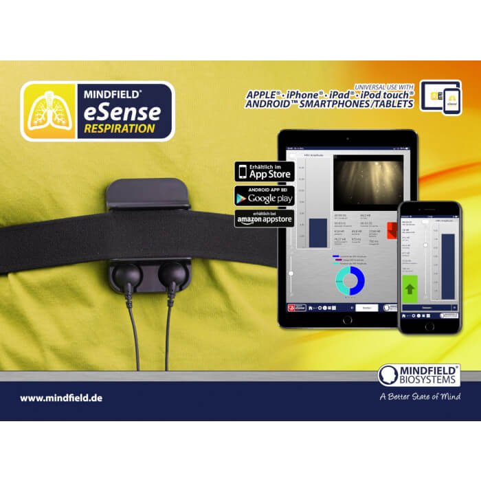
The HRV training biofeedback loop continuously monitors HRV parameters. It interprets physiological responses and adjusts training techniques based on real-time feedback. Sensors detect changes in HRV patterns and present them to the individual through visual or auditory cues. This feedback enables individuals to modulate their HRV responses, improving performance, stress management, and overall well-being.

In summary, while HRV training offers significant potential benefits, incorporating biofeedback guidance can enhance its effectiveness and efficiency. Biofeedback gives individuals real-time information about their physiological responses. This information allows them to adjust their techniques and practices for optimal results. The immediate feedback loop enhances awareness and control over HRV patterns, leading to more targeted and personalized interventions. This approach improves outcomes in stress management, athletic performance, and emotional regulation.

Moreover, biofeedback guidance ensures that individuals adhere to proper techniques and protocols, maximizing the therapeutic benefits of HRV training. Integrating biofeedback into HRV training enhances performance outcomes and deepens the understanding of the mind-body connection. It also enables individuals to modulate HRV patterns more effectively, improving health and performance.

Parameters of HRV Training with Biofeedback Guidance

Heart Rate Variability (HRV) training with biofeedback guidance monitors various parameters and indexes. This approach helps optimize training outcomes and health management. Understanding these parameters is crucial for assessing autonomic nervous system function and guiding interventions to improve HRV patterns.

The interval between successive normal heartbeats (between consecutive R waves on an electrocardiogram) tracing is called the NN interval. This interval is also referred to as the normal-to-normal interval. In other words, it measures the time elapsed from one heartbeat to the next normal heartbeat. The NN interval is the basic unit for calculating heart rate variability (HRV). It represents the heart’s rhythm in a stable state.

NN intervals are crucial for analyzing HRV because they reveal variations in the timing between heartbeats. This variation reflects the dynamic interplay of the autonomic nervous system. HRV analysis assesses the balance between sympathetic (fight-or-flight) and parasympathetic (rest-and-digest) nervous system activity. This measurement is based on evaluating differences between successive NN intervals. The balance between sympathetic and parasympathetic systems is vital for regulating various physiological functions and maintaining overall health.

Here are the key parameters and indexes of HRV training with biofeedback guidance.

1. Time Domain Parameters:

SDNN (Standard Deviation of NN intervals): SDNN measures the overall variability of NN intervals, reflecting both sympathetic and parasympathetic influences. A higher SDNN indicates greater HRV and better autonomic function. Typical range: 50-100 ms.

RMSSD (Root Mean Square of Successive Differences): RMSSD reflects parasympathetic activity and short-term HRV. Higher RMSSD values suggest better vagal tone and enhanced relaxation response. The normal range is 20-50 ms.

2. Frequency Domain Parameters:

LF (Low Frequency): LF power represents a mixture of sympathetic and parasympathetic influences, reflecting baroreceptor activity and sympathetic modulation. Increased LF may indicate heightened sympathetic activity or reduced parasympathetic activity. This change could suggest a state of heightened arousal, stress, or a diminished relaxation response. Typical range: 20-40% of total power.

HF (High Frequency): HF power primarily reflects parasympathetic activity and respiratory sinus arrhythmia. Higher HF indicates the prevalence of vagal tone and relaxation response. Elevated HF values suggest a dominance of parasympathetic nervous system activity, particularly the vagus nerve’s influence on the heart. A higher HF indicates a more robust relaxation response and better regulation of physiological functions associated with rest and digestion. Typical range: 40-60% of total power.

3. Nonlinear Parameters:

SD1 (Standard Deviation 1): SD1 represents short-term HRV and reflects beat-to-beat variability influenced by respiratory sinus arrhythmia. Higher SD1 values indicate better parasympathetic modulation, reflecting increased variability in heart rate patterns associated with the respiratory cycle. This mechanism suggests a more decisive influence of the parasympathetic nervous system on heart rate regulation. It promotes relaxation and adaptability to changing physiological demands. The typical range is 5-15 milliseconds.

SD2 (Standard Deviation 2): SD2 reflects long-term HRV and overall variability influenced by sympathetic and parasympathetic activity. Higher SD2 values suggest greater overall HRV, indicating increased variability in heart rate patterns over more extended periods. This broader variability is linked to better adaptability to stressors. It also shows improved cardiovascular health and enhanced overall well-being. The typical range for this variability is 20-50 milliseconds.

4. Others:

Coherence: Coherence measures the degree of synchronization between respiration and heart rate oscillations. Higher coherence values indicate better cardiovascular and respiratory coupling, reflecting more efficient autonomic function.

Unlike other HRV parameters, coherence measures 0 to 1 on a scale. Higher values indicate more robust synchronization between respiratory and heart rate rhythms.

A coherence value closer to 1 indicates high synchronization, reflecting efficient cardiovascular and respiratory coupling. This synchronization is associated with optimal autonomic nervous system function and cardiovascular health. On the other hand, a coherence value closer to 0 suggests weaker synchronization between respiratory and heart rate rhythms. This may indicate autonomic dysfunction or other health issues.

Coherence doesn’t have a specific range like other HRV parameters. Interpreting coherence values involves assessing the synchronization strength between respiratory and heart rate oscillations. This helps understand autonomic nervous system function and cardiovascular health.

Importance of HRV Training with Biofeedback Guidance Parameters in Performance and Health Management

Heart Rate Variability (HRV) training with biofeedback guidance is valuable for optimizing performance and health outcomes. By monitoring and modulating HRV parameters with real-time feedback, individuals can enhance their physiological resilience, emotional regulation, and overall well-being. 

Performance Optimization:

HRV training with biofeedback guidance provides athletes, performers, and professionals with a strategic approach to optimizing performance outcomes. Individuals can optimally balance sympathetic and parasympathetic nervous system activity by fine-tuning HRV parameters such as SDNN, RMSSD, LF, HF, SD1, SD2, and coherence. This balance fosters a state of physiological coherence, promoting peak performance, endurance, and recovery. By leveraging biofeedback to adjust real-time training protocols, individuals can adapt to changing demands, manage fatigue, and maximize their competitive edge.

Stress Resilience and Emotional Regulation:

Biofeedback-guided HRV training empowers individuals to manage stress better and regulate their emotions in high-pressure situations. Monitoring HRV parameters, such as LF, HF, SD1, and coherence, helps individuals understand their autonomic nervous system activity and stress response. Targeted interventions, like controlled breathing exercises and mindfulness techniques, allow individuals to modulate HRV patterns. These techniques induce relaxation, reduce anxiety, and enhance emotional resilience. Integrating biofeedback into HRV training boosts self-awareness and control over physiological and psychological responses. This integration fosters a more adaptive stress-coping mechanism.

Cardiovascular Health and Disease Prevention:

HRV training with biofeedback guidance holds promise for promoting cardiovascular health and mitigating the risk of heart disease. Individuals can enhance autonomic nervous system function and cardiovascular regulation by improving HRV parameters such as SDNN, RMSSD, LF, HF, SD1, SD2, and coherence. This optimization reduces the burden on the heart, lowers blood pressure, and improves vascular function. Incorporating biofeedback into HRV training provides personalized feedback on cardiovascular responses. This feedback allows individuals to tailor interventions to meet their specific needs. Over time, this proactive approach to cardiovascular health management may help prevent the onset of heart disease and related complications.

Sleep Quality Enhancement:

HRV training with biofeedback guidance can also improve sleep quality and health. Monitoring HRV parameters like RMSSD, HF, and coherence helps individuals assess autonomic nervous system activity and its effect on sleep patterns. Biofeedback-guided interventions, including relaxation techniques and breathing exercises, help individuals regulate their HRV patterns. These interventions promote relaxation and improve sleep onset and maintenance. Integrating HRV training with biofeedback into a bedtime routine can enhance sleep quality, reduce sleep disturbances, and improve overall sleep health.

Chronic Pain Management:

For individuals dealing with chronic pain conditions, HRV training with biofeedback guidance offers a complementary approach to pain management. Monitoring HRV parameters such as SDNN, LF, and coherence provides valuable insights into autonomic nervous system function. Furthermore, it clarifies the connection between this function and pain perception. Biofeedback-guided interventions, such as deep breathing exercises and mindfulness practices, help modulate HRV patterns. This modulation can reduce pain intensity and improve pain tolerance. Regular HRV training sessions with biofeedback guidance can potentially lead to a decreased reliance on pain medications. Additionally, these sessions may enhance functional abilities and improve overall quality of life.

Cognitive Function Enhancement:

In addition to physical health benefits, HRV training with biofeedback guidance can also support cognitive function and mental well-being. By monitoring HRV parameters such as SDNN, RMSSD, and coherence, individuals can assess their autonomic nervous system activity and its impact on cognitive performance.

Biofeedback-guided HRV training influences cognitive function through several physiological and psychological mechanisms. By providing real-time feedback on HRV parameters, individuals can effectively modulate autonomic nervous system activity. Consequently, this promotes a balanced response characterized by increased parasympathetic activity and decreased sympathetic activity. This physiological balance reduces stress levels and enhances cardiorespiratory efficiency, facilitating optimal oxygenation and blood flow to the brain. Biofeedback-guided HRV training may also synchronize brainwave patterns with HRV parameters, fostering coherence between brain and heart activity. 

Combined with consistent modulation of HRV patterns, this coherence promotes neuroplasticity and strengthens neural connections associated with cognitive function. Biofeedback-guided HRV training creates an optimal physiological state for cognitive functioning, improving attention, memory, decision-making, and overall mental well-being.

Biofeedback-guided interventions, such as cognitive-behavioral techniques and attentional focus exercises, can help modulate HRV patterns to enhance cognitive function, including attention, memory, and decision-making. By integrating HRV training with biofeedback guidance into a cognitive training regimen, individuals may experience improved cognitive resilience, enhanced mental clarity, and greater overall cognitive well-being.

Personalized Health Management:

HRV training with biofeedback guidance provides a personalized health management approach. It allows individuals to monitor and optimize physiological responses in real time. Tracking HRV parameters and adjusting training protocols help address specific health concerns like chronic pain, sleep disturbances, and mood disorders. This approach empowers individuals to engage actively in their health and well-being, improving adherence to therapeutic interventions and fostering long-term behavior change. Integrating HRV training with biofeedback into routine health practices can cultivate resilience, enhance performance, and optimize quality of life.

Overall Well-being Promotion:

Ultimately, HRV training with biofeedback guidance promotes overall well-being by empowering individuals to participate actively in their health and performance optimization. Monitoring HRV parameters and adjusting real-time training protocols help individuals cultivate resilience. Additionally, these adjustments reduce stress and enhance physical, emotional, and cognitive well-being. Biofeedback-guided HRV training supports a holistic approach to health management. It addresses the interconnectedness of mind and body and promotes sustainable behavior change. Incorporating HRV training with biofeedback guidance into daily routines enables individuals to unlock their full potential. Consequently, they can thrive in all aspects of life.

In conclusion, HRV training with biofeedback guidance parameters offers a comprehensive approach to performance enhancement and health management. Leveraging real-time feedback to modulate HRV patterns allows individuals to optimize their physiological responses. In addition, this modulation promotes resilience and supports overall health and well-being. As a result, individuals can achieve peak performance while maintaining their overall health. Through personalized interventions and targeted training protocols, HRV training with biofeedback guidance empowers individuals to lead healthier, more fulfilling lives.

How to Regulate and Optimize HRV Parameters and Patterns

Regulating and optimizing HRV parameters and patterns is crucial for enhancing overall health and well-being. By understanding how to modulate these parameters in both high and low cases, individuals can achieve a balanced autonomic nervous system response and promote optimal physiological function. Here are explored strategies for regulating and optimizing HRV parameters, including SDNN, RMSSD, LF, HF, SD1, SD2, and coherence, in various scenarios.

SDNN (Standard Deviation of NN Intervals):

  • High SDNN: To regulate high SDNN, individuals can engage in relaxation techniques such as deep breathing exercises, meditation, or progressive muscle relaxation. These practices promote parasympathetic activation, reducing sympathetic dominance and decreasing SDNN.
  • Low SDNN: Conversely, to increase low SDNN, individuals can incorporate aerobic exercise into their routine, engage in stress-reducing activities such as yoga or tai chi, or ensure adequate sleep and hydration. These interventions enhance overall HRV and increase SDNN.

RMSSD (Root Mean Square of Successive Differences):

  • High RMSSD: Techniques such as paced breathing, mindfulness meditation, or biofeedback-assisted relaxation can help increase RMSSD by enhancing parasympathetic activity and promoting vagal tone.
  • Low RMSSD: To address low RMSSD, individuals can focus on stress management strategies, ensure sufficient sleep quality and duration, and adopt a healthy lifestyle that includes regular physical activity and balanced nutrition.

LF (Low Frequency) and HF (High Frequency):

  • High LF: To regulate high LF, individuals can engage in relaxation techniques such as diaphragmatic breathing, progressive muscle relaxation, or guided imagery. These practices help reduce sympathetic activity and rebalance the autonomic nervous system.
  • Low LF: Conversely, to increase low LF, individuals can engage in moderate-intensity aerobic exercise, practice stress-inducing activities such as cold exposure or intermittent hypoxic training, or consume caffeine in moderation.
  • High HF: Techniques such as paced breathing, mindfulness meditation, or biofeedback-assisted relaxation can help increase HF by enhancing parasympathetic activity and promoting vagal tone.
  • Low HF: To address low HF, individuals can focus on stress management strategies, ensure sufficient sleep quality and duration, and adopt a healthy lifestyle that includes regular physical activity and balanced nutrition.

SD1 (Standard Deviation 1) and SD2 (Standard Deviation 2):

  • High SD1 and SD2: Techniques such as paced breathing, coherence training, or mindfulness meditation can help increase both SD1 and SD2 by promoting parasympathetic activation and enhancing heart rate variability.
  • Low SD1 and SD2: To address low SD1 and SD2, individuals can use relaxation techniques, stress management strategies, and regular physical activity to enhance overall HRV and improve autonomic function.

Coherence:

  • High Coherence: Techniques such as coherent breathing, HRV biofeedback training, or mindfulness-based practices can help increase coherence by promoting synchronization between respiratory and heart rate oscillations.
  • Low Coherence: To address low coherence, individuals can focus on improving relaxation skills, practicing slow, deep breathing exercises, and incorporating coherence training into their daily routines.

Implementing strategies tailored to specific HRV parameters and patterns helps individuals regulate their autonomic nervous system function. This regulation effectively optimizes their overall health, well-being, and performance. As a result, people see enhancements in all these areas.

Techniques for Managing HRV Parameters with Biofeedback-Guided Training

Breathing Techniques

Diaphragmatic Breathing:

Diaphragmatic breathing (see video explanation), also known as abdominal or deep breathing, involves engaging the diaphragm to take slow, deep breaths.

To practice diaphragmatic breathing, individuals should first sit or lie comfortably. Next, they should place one hand on their abdomen and inhale deeply through their nose, allowing their belly to rise. Afterward, they should exhale slowly through their mouth while contracting their abdominal muscles.

Diaphragmatic breathing involves slow, deep inhalations and exhalations that engage the diaphragm. This practice enhances HRV by promoting relaxation and activating the parasympathetic nervous system. As individuals continue diaphragmatic breathing, they stimulate the vagus nerve. This stimulation leads to increased vagal tone and more significant variability in heart rate. Consequently, individuals experience higher HRV, which indicates a more adaptive autonomic response and improved cardiovascular health.

Paced Breathing:

 Another effective technique is paced breathing (see video explanation), which entrains respiratory and heart rate rhythms to a specific frequency or pattern. Paced breathing involves synchronizing the breathing patterns with a particular rhythm or pace, typically guided by visual or auditory cues. Common-paced breathing techniques include coherent breathing (breathing at a rate of 6 breaths per minute) and resonance frequency breathing (see video explanation) (aligning breathing with individual resonance frequency). Paced breathing enhances HRV by promoting rhythmic heart rate patterns and coherence between respiratory and heart rate oscillations. These breathing techniques foster relaxation and stress reduction and optimize autonomic balance, ultimately improving HRV parameters and overall well-being.

Changes in HRV parameters during breathing techniques reflect the modulation of autonomic nervous system activity. Specifically, these changes include an increase in parasympathetic activity and a decrease in sympathetic activity. Additionally, breathing techniques enhance respiratory sinus arrhythmia and promote physiological coherence. As a result, parameters such as SDNN, RMSSD, HF, SD1, SD2, and Coherence increase. In contrast, LF decreases.

By practicing breathing techniques regularly, individuals can optimize HRV parameters, promote relaxation, and support overall health and well-being.

Exercise:

  • Aerobic Exercise: Aerobic exercise, such as walking, running, cycling, swimming, or dancing, involves continuous, rhythmic movements that increase heart rate and oxygen consumption. Regular aerobic exercise improves cardiovascular fitness, enhances respiratory efficiency, and increases HRV. To optimize HRV benefits, individuals should engage in moderate-intensity aerobic exercise for at least 30 minutes most days a week.
  • Strength Training: Strength training exercises, such as weightlifting, resistance band workouts, or bodyweight exercises, involve repetitive muscle contractions against resistance. While strength training primarily targets muscular strength and endurance, it can also indirectly improve HRV by promoting cardiovascular health and metabolic efficiency.

Moderate-intensity aerobic exercise increases HRV, particularly HF power. Regular physical activity can help rebalance autonomic nervous system activity by enhancing parasympathetic tone and reducing sympathetic dominance.

Mindfulness Practices:

  • Meditation: Meditation involves focusing on a specific object, thought, or sensation to cultivate present-moment awareness and mental clarity. Mindfulness meditation practices, including focused attention meditation, loving-kindness meditation, and body scan meditation, promote relaxation, reduce stress, and increase HRV by enhancing parasympathetic activation and emotional regulation.
  • Mindful Breathing: Mindful breathing, or breath awareness, involves paying attention to the sensations of breathing without judgment. By consciously observing each inhalation and exhalation, individuals can anchor their awareness in the present moment, reduce rumination, and enhance HRV by promoting relaxation and stress reduction.

Diet:

  • Balanced Nutrition: A balanced diet rich in fruits, vegetables, whole grains, lean proteins, and healthy fats supports overall health and can positively influence HRV. Certain nutrients, such as omega-3 fatty acids, magnesium, and antioxidants, have been associated with improved cardiovascular function and increased HRV. Conversely, excessive processed foods, sugary beverages, and high-fat meals may negatively impact HRV.

Sleep Hygiene:

  • Consistent Sleep Schedule: Maintaining a consistent sleep schedule by going to bed and waking up at the same time daily helps regulate circadian rhythms and supports healthy sleep patterns. Adults should aim for 7-9 hours of sleep daily to optimize HRV and overall well-being.
  • Sleep Environment: Creating a comfortable, relaxed, dark, and quiet environment promotes restful sleep and enhances HRV. To improve sleep quality, individuals should minimize exposure to electronic devices before bedtime, avoid caffeine and alcohol close to bedtime, and establish relaxing bedtime rituals.

Stress and Anxiety Management:

  • Stress Reduction Techniques: Stress reduction techniques, such as progressive muscle relaxation, guided imagery, or journaling, help alleviate tension and promote relaxation. By practicing stress management strategies regularly, individuals can reduce chronic stress levels, improve emotional resilience, and enhance HRV.
  • Anxiety Management Strategies: Anxiety management techniques, such as cognitive-behavioral therapy (CBT), deep breathing exercises, or mindfulness-based stress reduction (MBSR), empower individuals to cope effectively with anxious thoughts and emotions. These strategies promote emotional regulation, reduce physiological arousal, and increase HRV.

Implementing these techniques into biofeedback-guided HRV training can help individuals optimize HRV parameters, enhance autonomic nervous system function, and promote overall health and well-being.

Understanding the Difference Between HRV Training Devices and Biofeedback-Guided HRV Training Devices

Heart Rate Variability (HRV) training has gained popularity as a method for improving physiological resilience and overall well-being. While both HRV training devices and biofeedback-guided HRV training devices aim to enhance HRV patterns, there are fundamental differences between the two approaches.

HRV Training Devices:

HRV training devices typically measure and analyze HRV patterns without real-time feedback or intervention. These devices collect heart rate data over time and provide metrics such as SDNN, RMSSD, LF, HF, SD1, SD2, and coherence, which reflect autonomic nervous system activity and cardiovascular function. HRV training devices may include wearable heart rate monitors, smartphone apps, or standalone HRV measurement tools. 

Users of HRV training devices often review their HRV data retrospectively and adjust lifestyle factors, such as exercise, sleep, stress management, and nutrition, to optimize HRV patterns. While HRV training devices offer valuable insights into autonomic function, they lack real-time feedback mechanisms for guiding interventions and behavior modifications.

Biofeedback-Guided HRV Training Devices:

In contrast, biofeedback-guided HRV training devices combine HRV measurement with real-time feedback and intervention capabilities. These devices monitor HRV parameters and provide immediate feedback to users, allowing them to modulate their physiological responses in real-time. Biofeedback-guided HRV training devices typically employ visual, auditory, or tactile cues to indicate changes in HRV patterns and guide users in achieving desired physiological states. Users can actively influence their HRV patterns and promote physiological coherence by engaging in paced breathing, relaxation exercises, or mindfulness practices. 

Biofeedback-guided HRV training devices may include wearable sensors, smartphone apps, or standalone biofeedback devices equipped with HRV monitoring capabilities. Integrating real-time feedback into HRV training facilitates personalized interventions, enhances self-regulation skills, and promotes efficient physiological adaptation.

Key Differences:

The primary difference between HRV training devices and biofeedback-guided HRV training devices lies in providing real-time feedback and intervention capabilities. While HRV training devices focus on data collection and retrospective analysis, biofeedback-guided HRV training devices enable users to engage in HRV modulation through immediate feedback and intervention actively. Biofeedback-guided HRV training devices offer a dynamic and interactive approach to HRV training, empowering users to optimize autonomic function and enhance overall well-being in real time.

In summary, while HRV training devices and biofeedback-guided HRV training devices aim to improve HRV patterns, the latter approach distinguishes itself by incorporating real-time feedback. By providing immediate feedback and intervention capabilities, biofeedback-guided HRV training devices offer a more interactive and personalized approach to HRV modulation, facilitating efficient self-regulation and physiological adaptation.

Selecting the Right Biofeedback-Guided HRV Training Device

Choosing the appropriate biofeedback-guided HRV training device is essential for optimizing your HRV training experience and achieving your health and wellness goals. With various available options, consider several factors to ensure you select the device that best suits your needs and preferences.

Define Your Goals:

Before selecting a biofeedback-guided HRV training device, you must clarify your specific goals and objectives. Determine whether you aim to reduce stress, improve resilience, enhance athletic performance, manage chronic health conditions, or optimize overall well-being. Understanding your goals will help you narrow down the features and functionalities required in a training device.

Consider Device Features:

Please evaluate the features and capabilities of each biofeedback-guided HRV training device to ensure they align with your needs. Key features to consider include:
Real-time Feedback: Look for devices that offer immediate feedback on HRV parameters and guide you through real-time interventions.
User Interface: Assess the usability and intuitiveness of the device’s interface, including the display, controls, and navigation.
Customization Options: Choose a device for personalized settings and training programs tailored to your preferences and goals.
Compatibility: Ensure compatibility with your existing devices and platforms, such as smartphones, tablets, or computers, for seamless integration and data sharing.
Portability: Consider the device’s portability and convenience in various settings, including home, office, or travel.

Review Training Programs:

Explore the training programs and protocols each biofeedback-guided HRV training device offers to determine their suitability for your goals and preferences. Look for devices that provide a variety of guided interventions, such as paced breathing exercises, relaxation techniques, mindfulness practices, or coherence training. Consider whether the device offers customizable training programs to accommodate your needs and progression.

Assess Data Insights:

Examine the data insights and analytics each biofeedback-guided HRV training device provides to ensure they offer valuable information for monitoring progress and optimizing training outcomes. Look for devices that provide comprehensive metrics on HRV parameters, trends, patterns, and actionable recommendations for adjustments and improvements. Consider whether the device offers data visualization tools, reporting capabilities, or integration with third-party platforms for enhanced data analysis and interpretation.

Read Reviews and Recommendations:

Before making a purchase decision, research and read reviews from other users, healthcare professionals, or experts in the field. Pay attention to feedback regarding device reliability, accuracy, effectiveness, ease of use, and customer support. Seek recommendations from trusted sources or online communities to gain insights into the real-world experiences of users using the device for HRV training.

Budget Considerations:

Finally, consider your budget constraints and investment preferences when selecting a biofeedback-guided HRV training device. Compare the cost of each device relative to its features, capabilities, and long-term value for achieving your goals. Remember that while some devices may have a higher upfront cost, they may offer greater functionality, durability, and support for long-term use.

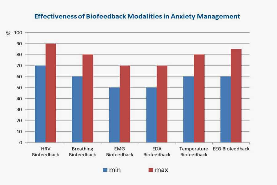
Enhancing Effectiveness with Combined Biofeedback Modalities:

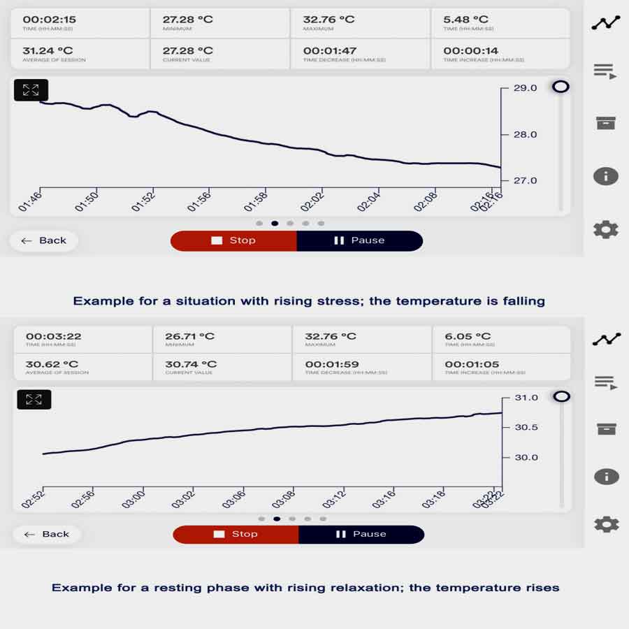
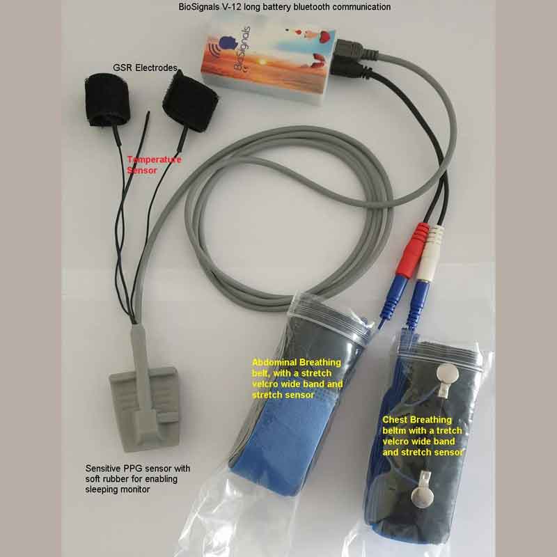
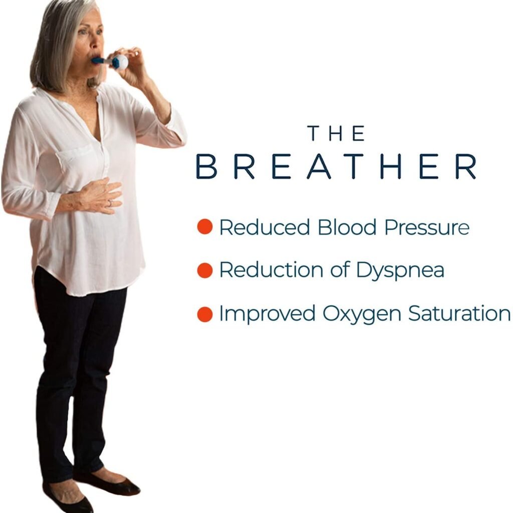
Combining biofeedback-guided HRV training with other biofeedback modalities, such as breathing biofeedback and galvanic skin response (GSR) biofeedback, can enhance the effectiveness of HRV training and optimize physiological regulation. Breathing biofeedback focuses on improving respiratory patterns and promoting relaxation through paced and diaphragmatic breathing techniques.

By incorporating breathing biofeedback into HRV training, individuals can enhance respiratory sinus arrhythmia, synchronize respiratory and heart rate rhythms, and achieve greater coherence between autonomic functions.

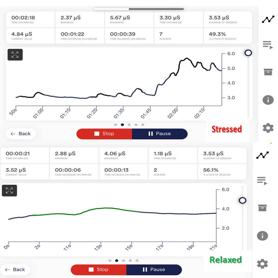
Similarly, GSR biofeedback measures changes in skin conductance, reflecting sympathetic nervous system arousal and emotional regulation. Integrating GSR biofeedback with HRV training allows individuals to monitor stress levels, enhance emotional awareness, and develop strategies for stress management. Combining multiple biofeedback modalities, individuals can access a comprehensive toolkit for self-regulation, optimizing HRV patterns, and promoting holistic well-being. This synergistic approach leverages the complementary benefits of different biofeedback modalities, resulting in more profound physiological and psychological effects.

Performing Biofeedback-Guided HRV Training Correctly

Performing biofeedback-guided HRV training according to recommendations is essential for maximizing its benefits and avoiding potential adverse or side effects. Here are guidelines and recommendations for conducting it effectively, including considerations for monitoring HRV parameters, scheduling training sessions, setting intensity levels, and implementing other best practices.

Attend to Proper Technique:

Ensure you use proper technique during biofeedback-guided HRV training sessions, especially when performing breathing exercises or relaxation techniques. Follow the instructions the device or app provides accurately and maintain a relaxed, comfortable posture throughout the session. Pay attention to your breathing pattern, heart rate variability, and any visual or auditory cues the device provides to guide your interventions.

Monitor HRV Parameters:

Monitor HRV parameters during training sessions regularly to track your progress and ensure you achieve the desired physiological responses. Focus on metrics such as SDNN, RMSSD, LF, HF, SD1, SD2, and coherence to assess changes in autonomic nervous system activity and cardiovascular function. Aim to maintain or increase HRV parameters within optimal ranges while avoiding excessive fluctuations that may indicate overexertion or stress.

Set Realistic Goals:

Establish realistic goals for your biofeedback-guided HRV training based on your needs, preferences, and health status. Start with achievable objectives, such as reducing stress levels, improving relaxation skills, or enhancing overall well-being, and gradually progress as you build proficiency and confidence in your training. Avoid setting overly ambitious goals that may lead to frustration or burnout.

Schedule Training Sessions:

Consistently schedule biofeedback-guided HRV training sessions into your daily or weekly routine to ensure regular practice and sustained progress. Allocate dedicated time for training sessions, ideally at the same time each day or week, to establish a consistent habit. Choose a quiet, comfortable environment free from distractions to optimize your focus and relaxation during training.

Adjust Intensity Levels:

Pay attention to your body’s responses and adjust the intensity levels of your training sessions accordingly. Start with gentle interventions and gradually increase the intensity as you become more comfortable and proficient. Monitor HRV parameters throughout the session and establish stop lines or thresholds to prevent overexertion or adverse reactions. Aim to balance challenging yourself and staying within your physiological limits.

Listen to Your Body:

Listen to your body’s signals and respect its limitations during biofeedback-guided HRV training. If you experience discomfort, fatigue, or negative symptoms such as dizziness or shortness of breath, pause the training session and take a break. Pay attention to any warning signs of overtraining or excessive stress and adjust your training intensity or duration accordingly.

Seek Professional Guidance:

Consider seeking guidance from a qualified healthcare professional or certified biofeedback therapist to ensure safe and effective implementation of biofeedback-guided HRV training. Consult with experts who can provide personalized recommendations, monitor your progress, and address any concerns or challenges you encounter during training. Professional guidance can enhance the effectiveness of your training and help you achieve optimal results while minimizing risks.

Maintain Balance and Consistency:

Maintain a balance between biofeedback-guided HRV training and other aspects of your lifestyle, including exercise, nutrition, sleep, and stress management. Avoid overemphasizing HRV training at the expense of other important health behaviors. Strive for consistency in your training regimen, but also allow for flexibility and adaptability to accommodate changes in your schedule or individual needs.

Following these guidelines and recommendations, you can perform biofeedback-guided HRV training correctly, maximize its benefits, and minimize potential adverse or side effects. Stay attentive to proper technique, monitor HRV parameters, set realistic goals, schedule training sessions consistently, adjust intensity levels appropriately, listen to your body, seek professional guidance, and maintain balance and consistency in your training approach. With diligent practice and mindful attention, biofeedback-guided HRV training can be a powerful tool for enhancing health, resilience, and well-being.

Conclusion

In conclusion, biofeedback-guided HRV training enhances physiological resilience, promotes stress management, and optimizes overall well-being. By leveraging real-time feedback mechanisms and interventions, individuals can actively modulate their autonomic nervous system activity, improve HRV patterns, and achieve greater physiological coherence.

Biofeedback-guided HRV training offers a dynamic and interactive method for self-regulation. It empowers individuals to cultivate skills for managing stress, enhancing relaxation, and promoting adaptive responses to environmental challenges. By incorporating techniques such as paced breathing, relaxation exercises, mindfulness practices, and coherence training, individuals can optimize HRV parameters, support cardiovascular health, and foster emotional well-being.

When performing biofeedback-guided HRV training, it is crucial to focus on proper technique. Additionally, individuals should monitor HRV parameters closely. Setting realistic goals is also essential. Consistently scheduling training sessions helps maintain progress. Adjusting intensity levels appropriately is necessary to match individual needs.

Furthermore, listening to the body’s signals ensures that the training remains effective. Seeking professional guidance as needed can provide additional support. Lastly, maintaining balance and consistency in the training approach is essential.

By following these guidelines and recommendations, individuals can fully harness the benefits of biofeedback-guided HRV training. As a result, they will be better equipped to achieve their health and wellness goals.

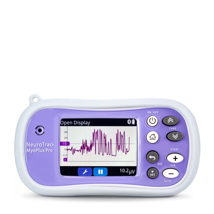
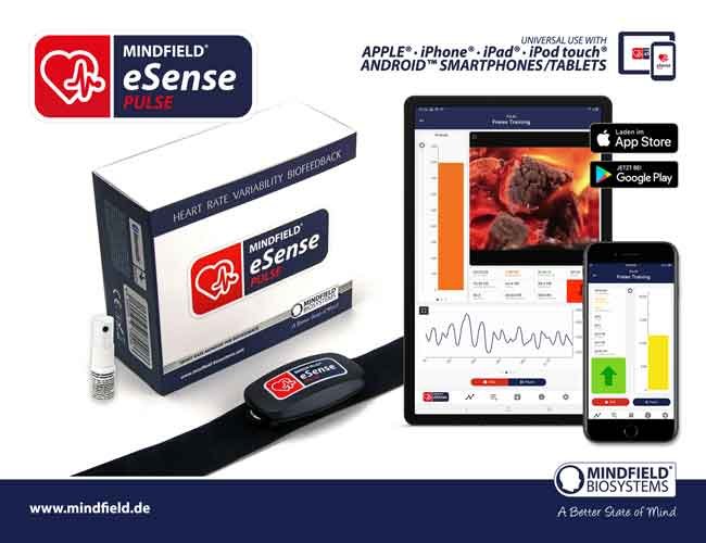
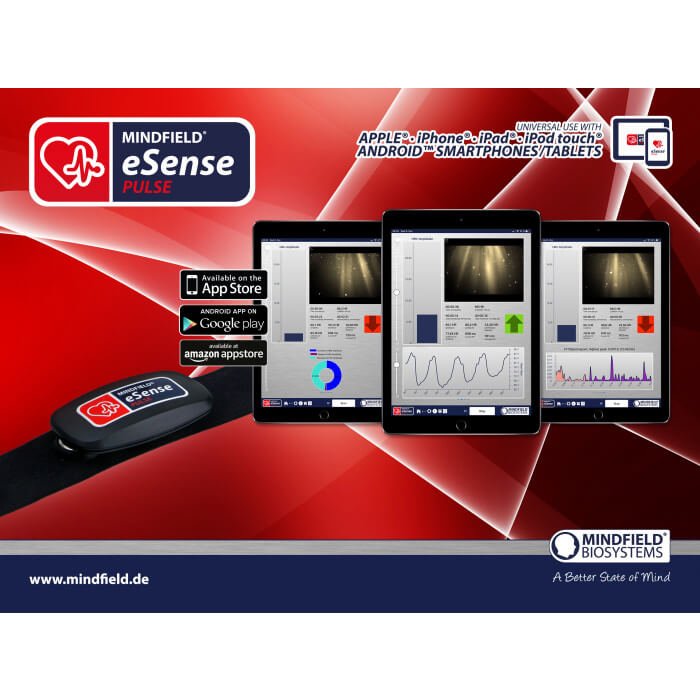
HRV Training Biofeedback Device

List of References

  1. Lehrer, P. M., & Gevirtz, R. (2014). Heart rate variability biofeedback: How and why does it work? Frontiers in Psychology, 5, 756.
  2. McCraty, R., Atkinson, M., et al. (2009). The coherent heart: Heart–brain interactions, psychophysiological coherence, and the emergence of system-wide order. Integral Review, 5(2), 10-115.
  3. Shaffer, F., & Ginsberg, J. P. (2017). An overview of heart rate variability metrics and norms. Frontiers in public health, 5, 258.
  4. Laborde, S., Mosley, E., et al. (2017). Heart rate variability and cardiac vagal tone in psychophysiological research–recommendations for experiment planning, data analysis, and data reporting. Frontiers in Psychology, 8, 213.
  5. McCraty, R., & Shaffer, F. (2015). Heart rate variability: new perspectives on physiological mechanisms, assessment of self-regulatory capacity, and health risk. Global advances in health and medicine, 4(1), 46-61.
  6. Laborde, S., & Allen, M. S. (2016). The role of cardiac vagal activity in the relationship between exercise and executive functioning. Brain and Cognition, 110, 102-106.
  7. Gevirtz, R. (2013). The promise of heart rate variability biofeedback: evidence-based applications. Biofeedback, 41(3), 110-120.
  8. Zaccaro, A., Piarulli, A., et al. (2018). How breath-control can change your life: A systematic review on psycho-physiological correlates of slow breathing. Frontiers in human neuroscience, 12, 353.
  9. van der Zwan, J. E., de Vente, W., et al. (2015). Physical activity, mindfulness meditation, or heart rate variability biofeedback for stress reduction: A randomized controlled trial. Applied Psychophysiology and Biofeedback, 40(4), 257-268.
  10. Grossman, P., & Taylor, E. W. (2007). Toward understanding respiratory sinus arrhythmia: Relations to cardiac vagal tone, evolution, and biobehavioral functions. Biological Psychology, 74(2), 263-285.
Secret of Success & Role of Biofeedback

Secret of Success & Role of Biofeedback

Unlocking the secret of success is a pursuit that captivates individuals across all walks of life. In this quest for excellence, biofeedback modalities serve as powerful tools. They offer valuable insights into the intricate interplay between mind and body. By harnessing the principles of biofeedback, individuals gain the ability to understand their physiological responses. Furthermore, they can regulate these responses effectively. This process helps them cultivate resilience and optimize performance in various domains.

Understanding Success. What is it? What psychophysiological parameters will contribute to being successful?

Success is a multifaceted concept that transcends mere accomplishment; it embodies realizing one’s goals and aspirations and fulfilling personal potential. Success is the attainment and satisfaction of an individual’s physical, mental, emotional, social, occupational, or personal goal or desire. In this context, we will delve into the essence of success, exploring what it truly means and identifying psychophysiological parameters that contribute to achieving it.

Defining Success

Success, in its truest sense, goes beyond material achievements and societal recognition. It encompasses a holistic and individualized perspective, acknowledging that the definition of success varies from person to person. Some may involve professional accomplishments, while others revolve around personal growth, relationships, or societal contributions. Understanding success requires a nuanced appreciation of individual values and passions and aligning one’s actions with one’s authentic self.

What Psychophysiological Parameters Contribute to Success?

Success is not solely a mental or physical endeavor but a dynamic interplay between the mind and body. Psychophysiological parameters are crucial in shaping an individual’s journey toward success. These parameters encompass a range of factors, including cognitive functions, emotional intelligence, and physiological responses. Exploring the connection between success’s psychological and physiological aspects unveils a deeper understanding of how our minds and bodies influence our ability to achieve our goals.

Cognitive Functions

The cognitive aspects of success involve mental processes such as perception, attention, memory, and problem-solving. A sharp, focused mind is better equipped to navigate challenges, make informed decisions, and persist in adversity. We will explore how cognitive functions contribute to success and how biofeedback modalities can enhance cognitive abilities.

Emotional Intelligence

Success often hinges on the ability to navigate complex social and emotional landscapes. Emotional intelligence, self-awareness, empathy, and effective interpersonal communication influence personal and professional success. Individuals with high emotional intelligence are better equipped to manage stress. Additionally, they build meaningful relationships and inspire others, which are vital to success. In this section, we will explore the role of emotional intelligence in achieving success. 

Physiological Responses

The body’s physiological responses, such as heart rate variability (HRV), electroencephalography (EEG) patterns, and galvanic skin response (GSR), offer valuable insights into our inner workings. These physiological indicators reflect our current state of arousal and stress and influence our cognitive and emotional functioning. Understanding and regulating these physiological responses can profoundly impact our ability to perform optimally and achieve success in various domains of life.

How Biofeedback Modalities Unveil the Secret of Success

Biofeedback modalities serve as powerful tools for uncovering the secrets of success by directly influencing and optimizing psychophysiological parameters. Through real-time feedback on various physiological processes, these innovative technologies enable individuals to gain both awareness and control over their mind-body interactions. Consequently, by harnessing biofeedback modalities, individuals can enhance cognitive functions, cultivate emotional intelligence, and regulate physiological responses. Ultimately, this integrated approach paves the way to unlocking the key to success.

Enhancing Cognitive Functions

Biofeedback modalities, such as neurofeedback, offer a unique opportunity to optimize cognitive functions by directly targeting brain activity. Through real-time monitoring of EEG patterns, individuals can learn to modulate their brainwaves to achieve heightened focus, creativity, and cognitive flexibility. By training the brain to operate more efficiently, biofeedback facilitates sharper mental understanding and enhanced problem-solving abilities, which are essential for success in various endeavors.

Cultivating Emotional Intelligence

Emotional intelligence, a cornerstone of success, can be cultivated and strengthened through biofeedback training. Techniques such as heart rate variability (HRV) biofeedback enable individuals to regulate their physiological responses to stress and emotions, fostering greater emotional resilience and self-awareness. By learning to maintain a balanced autonomic nervous system response, individuals can navigate challenging situations with composure, empathy, and clarity, fostering deeper connections and success in interpersonal relationships and leadership roles.

Regulating Physiological Responses

Biofeedback modalities regulate physiological responses like heart rate, respiration, and skin conductance. Through respiratory and GSR biofeedback, individuals can learn to induce relaxation, reduce stress, and enhance overall well-being. By achieving physiological coherence and balance, individuals can optimize their physical and mental functioning, paving the way for success in both personal and professional spheres.

By enhancing cognitive functions, cultivating emotional intelligence, and regulating physiological responses, biofeedback empowers individuals to unlock their full potential and achieve their goals with greater efficacy and fulfillment.

Illuminating Success with Electroencephalography (EEG) Biofeedback

Electroencephalography (EEG) biofeedback, also known as neurofeedback, unlocks the secret of success by harnessing the power of brainwave modulation. EEG biofeedback enables individuals to cultivate focused attention, creativity, and emotional balance by providing real-time feedback on brainwave activity. In academia, creative expression, and professional development, EEG biofeedback empowers individuals to optimize their cognitive functioning, unleash their creative potential, and achieve breakthrough success.

Enhancing Cognitive Functions

EEG biofeedback targets specific brainwave frequencies associated with cognitive functions such as attention, memory, and problem-solving. It trains individuals to increase or decrease activity in specific frequency bands, facilitating improved cognitive performance and mental acuity. In academic settings, it helps students strive for academic excellence. Meanwhile, it enables professionals to enhance productivity and innovation in corporate environments. Ultimately, EEG biofeedback offers a pathway to sharpened cognitive abilities and sustained success.

Cultivating Emotional Intelligence

Emotional intelligence, a critical component of success, is intricately linked to patterns of brainwave activity. EEG biofeedback enables individuals to modulate brainwave patterns linked to emotional processing. This modulation fosters greater emotional resilience and self-regulation. By cultivating a balanced emotional state, individuals enhance their empathic understanding. As a result, they can navigate interpersonal relationships with grace and authenticity. This ability ultimately unlocks the secret to success in leadership, teamwork, and personal fulfillment.

Facilitating Peak Performance

Peak performance is paramount in high-pressure environments such as competitive sports, performing arts, and executive leadership. EEG biofeedback offers athletes, artists, and professionals a means to enter flow states characterized by effortless focus, creativity, and peak performance. By training individuals to regulate their brainwave patterns associated with flow states, EEG biofeedback facilitates optimal performance. It unlocks the full potential of individuals striving for success in their fields.

Promoting Neuroplasticity and Personal Growth

One of the most remarkable aspects of EEG biofeedback is its ability to promote neuroplasticity, which refers to the brain’s capacity to reorganize and adapt in response to experience. By using targeted neurofeedback protocols, individuals can strengthen neural connections associated with desired cognitive and emotional states. As a result, they facilitate personal growth and self-actualization. Whether overcoming limiting beliefs, cultivating resilience in adversity, or fostering a growth mindset, EEG biofeedback empowers individuals. It helps them transcend barriers and unlock new levels of success in their lives.

Unlocking Success: Brainwave Optimization Strategies

Neurofeedback protocols can vary depending on individual goals and areas of focus related to success. However, some commonly used neurofeedback protocols that may contribute to success include:

SMR (Sensorimotor Rhythm) Training: 

SMR training focuses on enhancing sensorimotor rhythm activity in the brain, typically within the 12-15 Hz frequency range. By doing so, this protocol not only improves focus and attention but also boosts cognitive functioning. Consequently, these improvements are crucial in achieving success across academic, professional, and personal domains.

Alpha-Theta Training:

Alpha-theta training enhances alpha (8-12 Hz) and theta (4-8 Hz) brainwave activity, promoting deep relaxation and creativity. This protocol can reduce stress, increase emotional resilience, and facilitate insight and problem-solving abilities, all of which are conducive to success in various domains.

Beta Training:

Beta training specifically targets beta brainwave activity (15-30 Hz), which, in turn, is linked with alertness, concentration, and cognitive performance. As a result, by increasing beta activity, individuals may subsequently experience heightened mental acuity. Moreover, this enhancement often leads to improved focus and, consequently, enhanced productivity. Ultimately, these benefits can contribute significantly to academic, professional, and creative success.

4. Peak Alpha Frequency (PAF) Training:

PAF training focuses on increasing the individual’s peak alpha frequency, which is unique to each person, and correlates with cognitive functioning and overall brain health. By optimizing PAF, individuals may experience improved memory, attention, and mental flexibility, supporting success in cognitive tasks and problem-solving.

Connectivity Training:

Connectivity training aims to improve functional connectivity between different brain regions, enhancing communication and coordination within the brain’s neural networks. This protocol can help optimize information processing and promote efficient brain functioning. Additionally, it facilitates the integration of cognitive, emotional, and physiological processes. These elements are essential for success in various domains.

Selecting neurofeedback protocols should rely on individual assessment, goals, and preferences. Ideally, this process occurs under the guidance of a qualified neurofeedback practitioner to ensure effective and personalized treatment. Additionally, success in neurofeedback training often requires consistency, patience, and commitment to the process over time.

By enhancing cognitive functions, cultivating emotional intelligence, facilitating peak performance, and promoting neuroplasticity, EEG biofeedback empowers individuals to achieve their goals with greater efficacy and fulfillment. Whether striving for academic excellence, professional success, or personal growth, EEG biofeedback offers a pathway to unlock the full potential of the human mind and unleash success in every aspect of life.

Mendi Neurofeedback Headband for Health

Neuro VIZR for Mental Clarity and Focus

Unlocking the Secret of Success with Heart Rate Variability (HRV) Training

Heart Rate Variability (HRV) training is a beacon of hope in biofeedback modalities. It offers a profound understanding of the body’s autonomic nervous system and its crucial role in pursuing success. At its core, HRV reflects subtle variations in the time intervals between successive heartbeats. This variation serves as an invaluable indicator of physiological resilience and adaptability. Through HRV training, individuals embark on a transformative journey. They delve deep into the nuances of their physiology. As a result, they unlock untapped potential and achieve unparalleled success.

In the bustling arenas of athletics, where victory often hinges on split-second decisions and unwavering determination, HRV training emerges as a game-changer. By honing their HRV, athletes cultivate the resilience needed to push past physical limitations, maintain optimal performance under duress, and emerge victorious on the field of play. 

In the fast-paced world of corporate leadership, where success hinges on strategic insight, resilience in adversity, and the ability to inspire and motivate teams, HRV training offers a beacon of hope.

By optimizing their HRV, leaders cultivate emotional intelligence, enhance stress management skills, and foster a calm and composed demeanor amidst chaos. With insights from HRV training, leaders navigate turbulent waters with grace and poise, steering their organizations toward unprecedented success and growth.

Moreover, HRV training transcends the boundaries of specific domains, offering universal benefits that extend to all facets of life. Whether pursuing academic excellence, striving for personal fulfillment, or embarking on a journey of self-discovery, individuals find solace and empowerment in HRV training. 

HRV training represents a beacon of hope in pursuing success, offering individuals the keys to unlock their full potential and achieve greatness in every aspect of their lives.

Breathing Success: Respiratory Biofeedback Techniques

Respiratory biofeedback techniques offer a pathway to success through breathing patterns and autonomic nervous system balance regulation. By guiding individuals to achieve coherent breathing patterns, respiratory biofeedback facilitates relaxation, stress reduction, and enhanced mental clarity (video). In domains such as sports performance, mindfulness practice, and stress management, respiratory biofeedback empowers individuals to harness the power of breath to optimize performance, foster resilience, and achieve peak success.

Harnessing Success with Galvanic Skin Response (GSR) Biofeedback

Galvanic Skin Response (GSR) biofeedback offers unique insights into the body’s sympathetic nervous system activity and its influence on emotional arousal and stress. By monitoring changes in skin conductance, GSR biofeedback provides individuals with valuable feedback on their stress levels and emotional reactivity. In public speaking, interpersonal communication, and performance arts fields, GSR biofeedback equips individuals with the tools to manage stage fright, enhance charisma, and convey confidence, unlocking the secret of success in high-pressure situations.

Empowering Success with Muscle Biofeedback

Muscle (EMG) biofeedback provides individuals with insights into muscular tension and relaxation, offering a gateway to success in various domains. By monitoring electromyographic (EMG) signals, muscle biofeedback enables individuals to develop awareness of muscular tension patterns and learn to release tension effectively. Muscle biofeedback empowers individuals to optimize movement efficiency. It also helps prevent injuries and achieve peak performance. Muscle biofeedback unlocks the secret to success in various fields by enhancing bodily awareness and control. These include physical rehabilitation, sports training, and stress management.

From HRV training to EEG biofeedback, GSR biofeedback, respiratory biofeedback, and muscle biofeedback, each modality offers unique insights and techniques for optimizing performance, fostering resilience, and achieving peak success. By harnessing the power of biofeedback, individuals can unlock their full potential and embark on a journey toward greater fulfillment and achievement in every aspect of their lives.

Biofeedback Modalities Applications in Various Domains

Whether in sports performance enhancement, stress reduction and mental health, academic and professional success, or creative and artistic expression, biofeedback empowers individuals to unlock their full potential and achieve their goals with precision and excellence. By harnessing the power of biofeedback, individuals can transcend limitations, cultivate resilience, and embark on a journey toward greater fulfillment and success in every aspect of their lives.

Unveiling the Secret of Success in Sports Performance Enhancement

Sports performance enhancement represents a domain where the pursuit of success is paramount. Biofeedback modalities offer athletes unique opportunities to unlock their full potential and achieve peak performance. By harnessing techniques such as HRV training, EEG biofeedback, and muscle biofeedback, athletes can optimize their physical and mental abilities, cultivate resilience, and elevate their performance to new heights. Whether striving for victory on the field, track, or court, biofeedback empowers athletes to unlock the secret of success and achieve their athletic goals with precision and excellence.

Unlocking Success Through Stress Reduction and Mental Health

Managing stress and maintaining mental well-being are essential to success in today’s fast-paced world. Biofeedback modalities provide individuals with practical tools for stress reduction and mental health enhancement. Through techniques such as respiratory biofeedback and GSR biofeedback, individuals can learn to regulate their physiological responses to stress, cultivate emotional resilience, and achieve greater clarity of mind. By prioritizing mental health and well-being, individuals can unlock the secret of success in their personal and professional lives, fostering greater fulfillment and productivity.

Revealing the Secret of Academic and Professional Success

Academic and professional success hinge upon cognitive abilities, emotional intelligence, and effective stress management. Biofeedback modalities offer students and professionals valuable tools for optimizing performance and achieving goals. Through EEG biofeedback and HRV training, individuals can enhance focus, memory retention, and cognitive flexibility, improving academic and professional outcomes. Individuals can confidently navigate challenges and succeed in their chosen fields by cultivating emotional and stress resilience.

Unleashing Creativity and Artistic Expression

Creative and artistic expression represent domains where unlocking the secret of success is synonymous with tapping into one’s inner creativity and authenticity. Biofeedback modalities provide artists, musicians, and performers with tools to enhance creativity, overcome performance anxiety, and achieve flow states. Through techniques such as EEG and respiratory biofeedback, individuals can enter states of heightened creativity, presence, and inspiration, unleashing their artistic potential and achieving success in their creative endeavors.

From Conflict to Connection: Biofeedback’s Impact on Family Dynamics

Each biofeedback modality offers unique benefits for promoting successful family relationships, from enhancing emotional regulation and stress management to improving communication and fostering a sense of unity and understanding.

Heart Rate Variability Training

HRV training can promote harmony and resilience within family relationships. By optimizing HRV, individuals develop greater emotional regulation and stress management skills, allowing them to navigate conflicts and challenges with composure and empathy. Family members who engage in HRV training may experience improved communication, deeper connections, and a greater sense of unity and understanding within the family unit.

Electroencephalography (EEG) Biofeedback:

EEG biofeedback can enhance cognitive functioning and emotional intelligence within the family. By training individuals to regulate their brainwave activity, EEG biofeedback can improve focus, attention, and decision-making abilities, fostering more meaningful interactions and mutual support among family members. Additionally, EEG biofeedback may help family members manage symptoms of anxiety, depression, or ADHD, contributing to a healthier and more harmonious family dynamic.

GSR biofeedback

In the family context, GSR biofeedback helps promote emotional awareness and empathy by teaching members to recognize and regulate their physiological responses to stressors. Practicing GSR biofeedback together allows family members to communicate more effectively, manage conflicts constructively, and cultivate a supportive and nurturing home environment.

Respiratory biofeedback

Breathing biofeedback focuses on optimizing breathing patterns, which can profoundly impact emotional regulation and stress reduction within the family. Respiratory biofeedback teaches individuals to control their breathing and activate the body’s relaxation response. As a result, it promotes calmness, reduces tension, and fosters a sense of peace and tranquility in the family environment. When family members practice respiratory biofeedback, they often experience improved communication and enhanced intimacy. Consequently, this practice can lead to a greater sense of connection and harmony within the family.

Electromyographic Biofeedback

Muscle (EMG) biofeedback can be beneficial for managing physical and emotional stress within the family. By learning to relax tense muscles and release bodily tension, family members can alleviate anxiety symptoms, promote physical comfort, and enhance overall well-being. Muscle biofeedback techniques may also address chronic pain or tension headaches, allowing family members to support each other in managing health challenges and improving quality of life.

By incorporating biofeedback practices into family life, individuals can cultivate resilience, deepen connections, and create a supportive and nurturing environment where each member can thrive.

Biofeedback modalities offer a powerful pathway to unlocking the secret of success by empowering individuals to harness the innate potential of their minds and bodies. Integrating biofeedback into personal and professional development practices helps individuals cultivate self-awareness, resilience, and optimal performance. As a result, this approach leads to greater fulfillment and achievement in all aspects of life.

Navigating the Path to Success: A Guide to Choosing the Right Biofeedback Modality

When choosing the most appropriate biofeedback modality, consider individual goals, preferences, and focus areas. This approach ensures better success and alignment with personal needs.

Here are some recommendations to guide your selection process:

  1. Identify Your Goals: Clarify your specific objectives for using biofeedback. Are you aiming to improve cognitive performance, manage stress, enhance athletic performance, or cultivate emotional intelligence? Understanding your goals will help you choose the most relevant biofeedback modality.
  2. Assess Your Needs: Evaluate your current strengths and areas for improvement. Consider cognitive functioning, emotional resilience, physiological stress responses, and physical performance. Identifying your needs will guide you in selecting the biofeedback modality that effectively addresses these areas.
  3. Consider Modalities’ Suitability: Assess the suitability of different biofeedback modalities based on your preferences, lifestyle, and comfort level with technology. For example, wearable biofeedback devices or smartphone applications may be suitable if you prefer non-invasive techniques and real-time feedback.
  4. Evaluate Effectiveness: Research the effectiveness and evidence base of various biofeedback modalities for your goals. Look for studies, reviews, and testimonials supporting the efficacy of each modality in achieving success in your target domains.
  5. Try Multiple Modalities:  Experiment with different biofeedback modalities to find the ones that resonate with you and deliver the desired results. Stay open-minded and explore various techniques to discover what works best for your unique needs and preferences.
  6. Integrate Complementary Approaches: Consider integrating multiple biofeedback modalities or combining biofeedback with other complementary approaches, such as mindfulness meditation, cognitive-behavioral therapy, or physical exercise. 
  7. Monitor Progress and Adjust Accordingly: Regularly monitor your progress and adjust your biofeedback training regimen as needed. Pay attention to changes in cognitive functioning, emotional well-being, stress levels, and performance outcomes. 

Conclusion

By following these recommendations and carefully considering your goals, needs, preferences, and the evidence base, you can choose the most appropriate biofeedback modality to support your journey toward success. Remember that success is a multifaceted endeavor, and biofeedback modalities can serve as valuable tools to unlock your full potential and achieve your goals in various domains of life.

In conclusion, unlocking the journey of the secret of success with biofeedback modalities illuminates a path of profound self-discovery and personal growth. Through practices such as heart rate variability (HRV) training, EEG biofeedback, and others, individuals gain insights into their physiology, empowering them to cultivate resilience, manage stress, and optimize cognitive performance. As we embrace the transformative power of biofeedback, let us embark on a collective journey of empowerment and self-realization, guided by the belief that success is not merely an endpoint but a continual process of growth and fulfillment.

Discover Your Path to Success with Our Curated Selection

Embark on an enriching journey towards success with our handpicked selection of biofeedback devices available on our Shop page. Delve into diverse cutting-edge products carefully curated to cater to your unique needs and aspirations. Whether you’re striving to boost cognitive abilities, master stress management, or elevate athletic performance, our affiliate offerings encompass a range of innovative solutions to help you realize your full potential. Explore our Shop page today and discover the perfect biofeedback device to accompany you on your personal and professional growth path.

List of References

1. Lehrer, P. M., & Gevirtz, R. (2014). Heart rate variability biofeedback: how and why does it work? Frontiers in Psychology, 5, 756.
2. McCraty, R., & Shaffer, F. (2015). Heart Rate Variability: New Perspectives on Physiological Mechanisms, Assessment of Self-regulatory Capacity, and Health Risk. Global Advances in Health and Medicine, 4(1), 46–61.
3. Khazan, I. (2013). Biofeedback and the Modification of Performance Anxiety: A Review of the Literature. Journal of Cognitive Psychotherapy, 27(3), 231–246.
4. Hammond, D. C. (2006). What is Neurofeedback: An Update. Journal of Neurotherapy, 10(4), 25–36.
5. Tan, G., Shaffer, F., Lyle, R., et al. (2016). Evidence-Based Heart Rate Variability (HRV) Biofeedback: A Technique to Reduce Symptoms of Autonomic Dysfunction and Stimulate Self-Regulation. Applied Psychophysiology and Biofeedback, 41(2), 139–140.
6. Landers, D. M., & Petruzzello, S. J. (1994). The Influence of Electroencephalographic Biofeedback on Performance in Pre-elite Archers. Medicine & Science in Sports & Exercise, 26(4), 505–512.
7. Gruzelier, J. H. (2014). EEG-neurofeedback for optimizing performance. I: A review of cognitive and affective outcomes in healthy participants. Neuroscience & Biobehavioral Reviews, 44, 124–141.
8. Penzlien, L., Kersten, J.,et al. (2020). Neurofeedback as a Tool for Enhancing Performance in Surgery – A Systematic Review. Frontiers in Human Neuroscience, 14, 588447.
9. Prinsloo, G. E., Rauch, H. G. L., et al. (2011). Noakes, T. D., & Derman, W. E. (2011). The effect of short-duration heart rate variability (HRV) biofeedback on cognitive performance during laboratory-induced cognitive stress. Applied Cognitive Psychology, 25(5), 792–801.

Biofeedback Devices for Anxiety

Biofeedback Devices for Anxiety Overcome

In today’s fast-paced and often stress-inducing world, anxiety has become a prevalent concern affecting millions of individuals worldwide. Whether triggered by work pressures, personal challenges, or societal expectations, anxiety can significantly impact one’s quality of life, emotional well-being, and overall health. Fortunately, advancements in technology have paved the way for innovative solutions to help individuals better manage and alleviate anxiety symptoms – biofeedback devices for anxiety. 

In the realm of anxiety management, biofeedback devices stand out as promising tools. They offer real-time insights into physiological responses and empower individuals to regulate their anxiety levels effectively. In today’s fast-paced world, where stressors abound, and mental well-being is paramount, the emergence of biofeedback devices for anxiety represents a significant stride towards personalized, tech-enabled solutions.

Understanding Anxiety

Anxiety is a complex and multifaceted emotion that manifests differently from person to person. While it’s normal to experience occasional feelings of worry or nervousness, anxiety becomes problematic when it interferes with daily functioning. When anxiety affects quality of life, it signals a need for effective management strategies. Understanding the nature of anxiety is essential for effectively managing it, and biofeedback devices for anxiety can play a significant role in this process.

Anxiety is characterized by feelings of unease, worry, or fear about future events, situations, or outcomes. It can manifest physically, emotionally, and cognitively, leading to symptoms such as increased heart rate, sweating, restlessness, difficulty concentrating, and irritability. To delve deeper into the causes, types, and symptoms of anxiety, we invite you to explore our article titled “Neurofeedback for Anxiety.”

By gaining a deeper understanding of anxiety and its underlying mechanisms, individuals can empower themselves to explore practical strategies for managing and alleviating its impact on their lives. Biofeedback devices offer valuable tools for this purpose. They help monitor and regulate physiological indicators of anxiety. As a result, individuals can take a proactive approach to self-care and enhance their emotional well-being.

Role of Biofeedback in Anxiety Management

Biofeedback is a therapeutic technique that helps individuals gain awareness and control over their physiological responses. It achieves this through real-time monitoring of bodily functions. In the context of anxiety management, biofeedback proves to be a valuable tool. Specifically, it helps identify and regulate the physical manifestations of stress and anxiety. Understanding the role of biofeedback in anxiety management provides insights into its effectiveness as a complementary therapy.

Principles of Biofeedback

Biofeedback relies on operant conditioning, where individuals learn to modify their physiological responses through feedback provided by monitoring devices. Individuals can recognize patterns by observing real-time data on parameters such as heart rate, respiration, skin conductivity, skin temperature, muscle tension, and brain activity. They can then make conscious adjustments to achieve desired physiological states.

How Biofeedback Works for Anxiety

Anxiety often involves heightened physiological arousal, including increased heart rate, shallow breathing, cold hands, muscle tension, and rumination. Biofeedback devices provide objective feedback on these physiological markers, allowing individuals to identify signs of anxiety and implement relaxation techniques to counteract them. Through repeated practice and reinforcement, individuals can learn to regulate their physiological responses, reducing the intensity and frequency of anxiety symptoms over time.

Different Modalities of Biofeedback Devices for Anxiety Management

Within the realm of anxiety management, a diverse array of biofeedback devices has emerged. Each device offers unique modalities to assist individuals in monitoring and regulating their physiological responses. Biofeedback devices for anxiety include various technologies, such as heart rate variability (HRV) monitors, electrodermal activity (EDA) sensors, respiration rate monitors, muscle tension sensors, temperature monitors, and EEG neurofeedback devices. 

Each of these devices serves a specific purpose in aiding individuals with anxiety. They provide real-time feedback on physiological parameters associated with stress and relaxation. By harnessing these biofeedback modalities, individuals gain greater awareness of their bodily responses to anxiety triggers. Consequently, they develop effective strategies for self-regulation and stress reduction.

Heart Rate Variability (HRV) Biofeedback Devices for Anxiety

HRV biofeedback focuses on optimizing the variability in the intervals between heartbeats. This practice promotes a state of physiological coherence associated with relaxation and emotional balance. HRV biofeedback devices for anxiety train individuals to regulate their heart rate variability, which refers to the variation in time intervals between successive heartbeats. 

The devices provide real-time feedback on heart rate patterns, typically through visual or auditory cues. Consequently, individuals learn to modulate their physiological responses. This process helps them achieve a state of coherence between heart rate variability and respiration.

Here’s how HRV biofeedback works in anxiety, including the initial changes and effects after a biofeedback session.

1. Initial Changes in Anxiety:

  • When individuals experience anxiety, their body’s autonomic nervous system becomes activated. This activation leads to physiological changes, such as an increased heart rate, shallow breathing, and heightened sympathetic arousal.
  • These physiological responses are often accompanied by decreased heart rate variability, reflecting a more rigid and less adaptive autonomic nervous system functioning.
  • Individuals may experience palpitations, chest tightness, sweating, nervousness, or dread.

2. During HRV Biofeedback Session:

  • During an HRV biofeedback session, individuals are typically instructed to focus on breathing. They also monitor their heart rate variability using biofeedback equipment.
  • Visual or auditory cues provide real-time feedback on changes in heart rate variability. These cues help individuals establish a coherent pattern between heart rate variability and respiration.
  • Through paced breathing exercises and relaxation techniques, individuals learn to synchronize their breathing with changes in heart rate variability. This synchronization promotes a shift towards parasympathetic dominance and reduces sympathetic arousal.
  • As individuals practice HRV biofeedback, they may experience a sense of relaxation, calmness, and increased awareness of their physiological responses.

Video – HRV Biofeedback: Breathing and HRV synchronization

3. After HRV Biofeedback Session:

  • After completing an HRV biofeedback session, individuals may experience immediate reductions in anxiety symptoms. For example, they might notice a decreased heart rate, reduced muscle tension, and an improved mood.
  • With continued practice over multiple sessions, individuals may notice longer-lasting effects. For instance, they might experience improvements in overall stress resilience, emotional regulation, and their ability to cope with anxiety triggers.
  • Over time, HRV biofeedback training can lead to enduring changes in autonomic nervous system functioning. This enhancement improves flexibility and adaptability in response to stressors.
  • Individuals may also develop greater self-awareness and self-regulation skills, which can help them manage anxiety more effectively in daily life.

The HRV biofeedback device for anxiety trains individuals to regulate their heart rate variability. This process promotes coherence between heart rate variability and respiration. Through repeated practice, HRV biofeedback can lead to immediate reductions in anxiety symptoms. Additionally, it can result in long-term improvements in stress resilience and emotional well-being.

Anxiety Management with Breathing Biofeedback Devices

Breathing Biofeedback involves paced breathing exercises to promote relaxation and reduce respiratory rate, leading to decreased sympathetic nervous system activity and increased parasympathetic activation.

Respiration biofeedback devices for anxiety is a technique that focuses on training individuals to control their breathing patterns to promote relaxation, reduce stress, and alleviate symptoms of anxiety. Here’s how breathing Biofeedback works in anxiety, including the initial changes and effects after a biofeedback session.

1. Initial Changes in Anxiety:

  • Anxiety often leads to physiological changes in the body, including increased respiratory rate, shallow breathing, and chest tightness.
  • Individuals may experience symptoms such as hyperventilation, where they breathe rapidly and shallowly, leading to decreased carbon dioxide levels in the blood and an imbalance in the body’s oxygen-carbon dioxide ratio.
  • These changes in breathing patterns can further exacerbate feelings of anxiety, leading to a cycle of increased stress and physiological arousal.

2. During Breathing Biofeedback Session:

  • During a breathing biofeedback session, individuals are typically connected to biofeedback equipment that monitors their respiration rate and depth.
  • Visual or auditory cues provide real-time feedback on changes in breathing patterns, allowing individuals to observe and adjust their respiration rate accordingly.
  • Individuals are guided to engage in paced breathing exercises, synchronizing their breathing with the cues provided by the biofeedback device.
  • By practicing slow, deep diaphragmatic breathing (link to the video: Diaphragmatic Breathing: How to Perform), individuals learn to regulate their respiratory rate, increase carbon dioxide levels in the blood, and promote relaxation.

Video – Exercise your proper breathing 4/6 with video-guide

3. After Breathing Biofeedback Session:

  • After completing a breathing biofeedback session, individuals may experience immediate reductions in anxiety symptoms, such as decreased heart rate, muscle tension, and feelings of stress.
  • By engaging in slow, deep diaphragmatic breathing, individuals activate the parasympathetic nervous system, which promotes relaxation and counters the sympathetic arousal associated with anxiety.
  • With continued practice over multiple sessions, individuals may notice longer-lasting effects, including improvements in overall respiratory function, stress resilience, and emotional well-being.
  • Breathing biofeedback training can also help individuals develop greater awareness of their breathing patterns and the ability to self-regulate their responses to stressors in daily life.

In summary, respiratory biofeedback devices for anxiety work by training individuals to control their breathing patterns. They also promote relaxation and reduce symptoms of anxiety. Through repeated practice, breathing biofeedback can lead to both immediate and long-term improvements. For instance, individuals may see enhancements in respiratory function, stress resilience, and emotional well-being. Ultimately, this training empowers individuals to manage anxiety more effectively.

EMG Biofeedback Devices for Anxiety Management

EMG Biofeedback utilizes surface electromyography (sEMG) to measure muscle tension levels. By becoming aware of and reducing excessive muscle tension, individuals can alleviate physical symptoms of anxiety, such as headaches and muscle stiffness.

EMG (electromyography) biofeedback devices for anxiety management are a technique that trains individuals to recognize and regulate muscle tension levels in their bodies. Here’s how EMG biofeedback works in anxiety, including the initial changes in anxiety and the effects after a biofeedback session.

1. Initial Changes in Anxiety:

  • Anxiety often manifests in the body as increased muscle tension, particularly in areas such as the neck, shoulders, jaw, and back.
  • Individuals may experience symptoms such as muscle tightness, clenched jaw, stiff neck, or tension headaches as a result of heightened stress and anxiety.
  • Chronic muscle tension can contribute to feelings of discomfort, fatigue, and physical stress, exacerbating the overall experience of anxiety.

2. During EMG Biofeedback Session:

  • During an EMG biofeedback session, individuals are connected to biofeedback equipment that measures their muscle activity, typically using surface electrodes placed on target muscle groups.
  • Visual or auditory cues provide real-time feedback on changes in muscle tension levels, allowing individuals to observe and learn to control their muscle activity.
  • Individuals are guided through relaxation techniques, such as progressive muscle relaxation or guided imagery, while their muscle tension is monitored on the biofeedback display.
  • Individuals learn to modulate their physiological responses and promote relaxation by consciously relaxing tense muscles and reducing muscle activity.

3. After EMG Biofeedback Session:

  • After completing an EMG biofeedback session, individuals may experience immediate reductions in muscle tension and feelings of physical relaxation.
  • By learning to release tension in specific muscle groups, individuals can alleviate physical symptoms associated with anxiety, such as muscle tightness or stiffness.
  • With continued practice over multiple sessions, individuals may notice longer-lasting effects, including improvements in overall muscle awareness, stress resilience, and emotional well-being.
  • EMG biofeedback training can also help individuals develop greater mindfulness of their muscle tension patterns and the ability to intervene proactively to prevent tension buildup in response to stressors.

In summary, EMG biofeedback devices for anxiety work by training individuals to recognize and regulate muscle tension levels. This process promotes relaxation and reduces the physical symptoms of anxiety. EMG biofeedback can lead to immediate and long-term improvements through repeated practice.

For instance, individuals may experience better muscle relaxation, increased stress resilience, and enhanced emotional well-being. Ultimately, this training empowers individuals to manage anxiety-related muscle tension more effectively in daily life.

Electrodermal Skin Activity (EDA) Biofeedback Devices

Electrodermal Skin Activity (EDA) or Galvanic Skin Response (GSR) biofeedback is a technique that trains individuals to recognize and regulate changes in skin conductance. Skin conductance reflects fluctuations in sympathetic nervous system activity and arousal levels. By learning to modulate skin conductance, individuals can effectively reduce physiological arousal and anxiety.

Here’s how EDA biofeedback works in anxiety management: Initially, individuals may notice changes in their anxiety levels as they begin to use the biofeedback device. After a biofeedback session, they often experience improvements in their ability to manage anxiety, including reduced arousal and a greater sense of calm.

1. Initial Changes in Anxiety:

  • Anxiety often leads to activation of the sympathetic nervous system, which can result in changes in skin conductance.
  • Skin conductance reflects the activity of sweat glands, which are controlled by the sympathetic nervous system, when individuals experience stress or anxiety, sweat gland activity increases, increasing skin conductance.
  • These changes in skin conductance can manifest as sweating, clamminess, or changes in skin temperature, which are physiological responses to sympathetic arousal.

2. During EDA Biofeedback Session:

  • During an EDA biofeedback session, individuals are connected to biofeedback equipment that measures their skin conductance level, typically using electrodes placed on the fingers or palms.
  • Visual or auditory cues provide real-time feedback on changes in skin conductance, allowing individuals to observe their physiological responses and learn to modulate them.
  • Individuals are guided through relaxation techniques, such as deep breathing, visualization, or mindfulness exercises, while the biofeedback display monitors their skin conductance.
  • Individuals learn to lower their skin conductance levels and promote a sense of calm by consciously inducing relaxation and reducing sympathetic arousal.

3. After EDA Biofeedback Session:

  • After completing an EDA biofeedback session, individuals may experience immediate reductions in skin conductance and feelings of physical relaxation.
  • By learning to regulate sympathetic nervous system activity, individuals can alleviate physiological symptoms associated with anxiety, such as sweating or clamminess.
  • With continued practice over multiple sessions, individuals may notice longer-lasting effects, including improvements in overall stress resilience, emotional well-being, and the ability to manage anxiety-related physiological responses.
  • EDA biofeedback training can also help individuals develop greater awareness of their physiological arousal patterns and the ability to intervene proactively to reduce stress and anxiety in daily life.

In summary, EDA biofeedback devices for anxiety work by training individuals to recognize and regulate changes in skin conductance. These changes reflect sympathetic nervous system activity. Through repeated practice, EDA biofeedback can lead to both immediate and long-term improvements. For example, individuals may experience enhancements in physiological relaxation, stress resilience, and emotional well-being. Ultimately, this training empowers individuals to manage anxiety-related physiological responses more effectively.

Temperature Biofeedback Devices for Anxiety Management

Temperature biofeedback is a technique that involves training individuals to regulate their skin temperature, typically in the hands or fingers, to promote relaxation and reduce symptoms of anxiety. Here’s how temperature biofeedback devices for anxiety management work in anxiety, including the initial changes in anxiety and the effects after a biofeedback session.

1. Initial Changes in Anxiety:

  • Anxiety often leads to physiological changes in the body, including alterations in peripheral blood flow and skin temperature.
  • When individuals experience stress or anxiety, the sympathetic nervous system becomes activated, leading to vasoconstriction in peripheral blood vessels, including those in the hands.
  • This vasoconstriction reduces blood flow to the hands and can decrease skin temperature, resulting in sensations of coldness or clamminess in the hands.
  • These changes in skin temperature are often accompanied by tension, discomfort, or physiological arousal associated with anxiety.

2. During Temperature Biofeedback Session:

  • During a temperature biofeedback session, individuals are connected to biofeedback equipment that measures their skin temperature, typically using thermistors or infrared sensors placed on the fingers or hands.
  • Visual or auditory cues provide real-time feedback on changes in skin temperature, allowing individuals to observe their physiological responses and learn to modulate them.
  • Individuals are guided through relaxation techniques, such as deep breathing, progressive muscle relaxation, or guided imagery, while the biofeedback display monitors their skin temperature.
  • By consciously inducing relaxation and promoting peripheral vasodilation, individuals learn to increase blood flow to the hands and raise skin temperature, fostering a sense of warmth and relaxation.

3. After Temperature Biofeedback Session:

  • After completing a temperature biofeedback session, individuals may experience immediate reductions in skin temperature and feelings of physical relaxation.
  • By learning to regulate peripheral blood flow and skin temperature, individuals can alleviate anxiety-related physiological symptoms, such as cold hands or clamminess.
  • With continued practice over multiple sessions, individuals may notice longer-lasting effects, including improvements in overall stress resilience, emotional well-being, and the ability to manage anxiety-related physiological responses.
  • Temperature biofeedback training can also help individuals develop greater awareness of their physiological arousal patterns and the ability to intervene proactively to reduce stress and anxiety in daily life.

In summary, temperature biofeedback devices for anxiety work by training individuals to regulate their skin temperature. This regulation promotes relaxation and reduces symptoms of anxiety. Temperature biofeedback can lead to immediate and long-term improvements through repeated practice. For example, individuals may experience better physiological relaxation, increased stress resilience, and enhanced emotional well-being. Ultimately, this training empowers individuals to manage anxiety-related physiological responses more effectively.

EEG Biofeedback Devices for Anxiety Management

EEG (Electroencephalography) biofeedback, also known as neurofeedback or EEG neurofeedback, is a technique that focuses on training individuals to regulate their brainwave activity to promote relaxation, reduce stress, and alleviate anxiety symptoms. For further information on the EEG Biofeedback (Neurofeedback) technique (including neurofeedback protocols) in anxiety management, please refer to the article titled “Neurofeedback for Anxiety Disorders.”
Here’s how EEG biofeedback devices for anxiety management work, including the initial changes in anxiety and the effects after a biofeedback session.

1. Initial Changes in Anxiety:

  • Anxiety often involves dysregulation of brainwave activity, including increased activity in regions of the brain associated with heightened arousal and emotional reactivity.
  • Individuals experiencing anxiety may exhibit patterns of excessive beta wave activity, which is associated with alertness, rumination, and stress.
  • Other individuals may show alpha, theta, or delta wave activity imbalances, which can contribute to difficulties in attention, relaxation, and emotional regulation.
  • These patterns of brainwave activity can exacerbate feelings of anxiety, leading to a cycle of heightened stress and physiological arousal.

2. During EEG Biofeedback Session:

  • During an EEG biofeedback session, individuals are connected to biofeedback equipment that measures their brainwave activity, typically using electrodes placed on the scalp (Video – Practical guides for measurement for EEG 10-20 system electrode placement for Neurofeedback).
  • Visual or auditory cues provide real-time feedback on changes in brainwave activity, allowing individuals to observe their brainwave patterns and learn to modulate them.
  • Individuals are guided through relaxation techniques, such as deep breathing, mindfulness, or visualization exercises, while their brainwave activity is monitored on the biofeedback display.
  • By consciously inducing a state of relaxation and promoting desirable patterns of brainwave activity, individuals learn to regulate their neural responses. This approach helps reduce stress and anxiety.

3. After EEG Biofeedback Session:

  • After completing an EEG biofeedback session, individuals may experience immediate reductions in anxiety symptoms, such as decreased rumination, improved mood, and increased feelings of calmness.
  • By learning to modulate their brainwave activity, individuals can alleviate both physiological and cognitive symptoms associated with anxiety. This modulation promotes a sense of emotional well-being and relaxation.
  • With continued practice over multiple sessions, individuals may notice longer-lasting effects, including improved brainwave regulation, stress resilience, and emotional self-regulation.
  • EEG biofeedback training can also help individuals develop greater self-awareness of their cognitive and emotional states. Additionally, it enhances their ability to intervene proactively. As a result, individuals can reduce stress and anxiety more effectively in daily life.

In summary, EEG biofeedback devices for anxiety management work by training individuals to regulate their brainwave activity. This regulation promotes relaxation and reduces symptoms of anxiety. EEG biofeedback can lead to immediate and long-term improvements through repeated practice. For instance, individuals may experience enhanced brainwave regulation, increased stress resilience, and improved emotional well-being. Ultimately, this training empowers individuals to manage anxiety-related symptoms more effectively.

Integrative Biofeedback Systems for Anxiety Management

  • Some biofeedback devices integrate multiple sensors and modalities to simultaneously provide comprehensive feedback on various physiological parameters.
  • These integrative systems may combine heart rate measurements, respiration, skin conductance, temperature, and muscle tension to offer a holistic assessment of the individual’s physiological state.
  • Integrative biofeedback systems can be beneficial for addressing complex physiological arousal patterns and tailoring interventions to individual needs.

By exploring the different types of biofeedback devices available, individuals can identify the most suitable tools for monitoring and regulating their physiological responses to anxiety. Biofeedback devices offer versatile options for promoting self-awareness, relaxation, and emotional well-being, whether focusing on heart rate variability, electrodermal activity, respiration, muscle tension, or brainwave activity.

Factors to Consider When Choosing a Biofeedback Device

Selecting a suitable biofeedback device is crucial for effectively managing anxiety and achieving desired outcomes. Several key factors should be considered to ensure compatibility with individual needs, preferences, and goals.

Accuracy and Reliability

  • Accuracy: Choose biofeedback devices that provide precise and reliable measurements of physiological parameters, as inaccurate data may compromise the effectiveness of biofeedback interventions.
  • Reliability: Look for devices validated through scientific research and testing to ensure consistent performance and reproducible results.

Ease of Use

  • User-Friendly Interface: Choose biofeedback devices with intuitive interfaces and clear instructions for operation, making it easy for individuals to navigate and utilize the device effectively.
  • Setup and Calibration: Consider the ease of setup and calibration procedures, as cumbersome or complex processes may deter users from using the device regularly.

Compatibility and Connectivity

  • Device Compatibility: Ensure the biofeedback device is compatible with the platforms and devices you intend to use, such as smartphones, tablets, or computers.
  • Connectivity Options: Look for biofeedback devices that offer multiple connectivity options, such as Bluetooth, USB, or wireless connectivity, to facilitate data transfer and integration with other devices and software.

Feedback Mechanisms

  • Visual Feedback: Evaluate the type and quality of visual feedback provided by the biofeedback device, such as graphs, charts, or animations, to ensure clarity and effectiveness in conveying physiological information.
  • Auditory Feedback: Consider whether the device offers auditory cues or sound effects to complement visual feedback, as auditory stimuli can enhance engagement and attention during biofeedback training sessions.

Customization and Personalization

  • Customization Options: Choose biofeedback devices that allow you to customize settings, parameters, and feedback thresholds to accommodate individual preferences and adapt to evolving needs.
  • Personalized Feedback: Look for biofeedback devices that offer customized feedback based on individual baseline measurements and progress, as personalized interventions are often more effective and motivating.

Cost Considerations

  • Initial Investment: Assess the upfront cost of purchasing the biofeedback device, including any additional accessories or software required for operation.
  • Long-Term Value: Consider the biofeedback device’s long-term value proposition, taking into account factors such as durability, warranty coverage, and potential for future upgrades or expansions.
  • Cost-Benefit Analysis: Evaluate the potential benefits of using the biofeedback device concerning its cost, weighing the investment against the expected outcomes and improvements in anxiety management and overall well-being.

By carefully considering these factors when choosing a biofeedback device, individuals can select the most suitable tool for their needs and maximize the effectiveness of their anxiety management efforts. Whether prioritizing accuracy, ease of use, connectivity, feedback mechanisms, customization, or cost-effectiveness, the suitable biofeedback device can empower individuals to gain greater self-awareness, regulate their physiological responses, and achieve meaningful progress in anxiety reduction and emotional resilience.

Essential Points to Remember

When selecting a biofeedback modality for managing anxiety, it’s crucial to consider the individual’s specific symptoms and underlying physiological responses. Different biofeedback modalities target various aspects of the body’s stress response system. Therefore, choosing the right modality can enhance its effectiveness in addressing particular symptoms associated with anxiety. For example, if an individual experiences prominent symptoms of rumination characterized by persistent and intrusive thoughts, EEG biofeedback may be particularly beneficial. By training individuals to regulate their brainwave activity, EEG biofeedback helps reduce excessive beta wave activity associated with rumination and promotes a state of calmness and cognitive clarity. 

Conversely, if an individual exhibits symptoms of tachycardia or an elevated heart rate, which indicate heightened sympathetic arousal, HRV (Heart Rate Variability) biofeedback may be more effective. HRV biofeedback helps by teaching individuals to modulate their heart rate variability. This approach promotes relaxation, reduces physiological arousal, and aids in restoring balance to the autonomic nervous system. Similarly, individuals experiencing symptoms such as muscle tension, sweating, or clamminess may benefit from EMG (Electromyography) or temperature biofeedback, which focuses on regulating muscle tension or peripheral blood flow, respectively. 

By tailoring the biofeedback modality to the individual’s specific symptoms and physiological responses, clinicians can maximize the effectiveness of biofeedback training in anxiety management and help individuals achieve meaningful improvements in their overall well-being.

How to Use Biofeedback Devices for Anxiety Relief

Biofeedback devices offer individuals a powerful tool for managing anxiety by providing real-time feedback on physiological responses and facilitating self-regulation techniques. Implementing biofeedback effectively involves understanding how to use these devices in a structured and purposeful manner to achieve anxiety relief.

Setting Realistic Goals

  • Start by setting specific, measurable, and achievable goals for using biofeedback to manage anxiety. Identify areas of concern or particular symptoms you want to address, such as reducing heart rate, decreasing muscle tension, or promoting relaxation.
  • Break down larger goals into smaller, manageable steps to track progress and maintain motivation. Celebrate achievements along the way to reinforce positive behaviors and outcomes.

Establishing Baselines

  • Before beginning biofeedback training, establish baseline measurements of relevant physiological parameters, such as heart rate, respiration rate, skin conductivity, and muscle tension. These baseline measurements serve as a reference point for monitoring progress and evaluating the effectiveness of interventions.
  • Conduct baseline assessments in a relaxed and calm state to obtain accurate baseline readings. Record baseline measurements regularly to track changes over time and adjust biofeedback strategies accordingly.

Practicing Regularly

  • Consistent practice is essential for maximizing the benefits of biofeedback training for anxiety relief. Incorporate biofeedback sessions into your daily routine and allocate dedicated time for practice.
  • Start with shorter practice sessions and gradually increase the duration and frequency as you become more comfortable with the techniques. To experience meaningful benefits, aim for regular practice sessions lasting at least 10-20 minutes.
  • Practice mindfulness and relaxation techniques during biofeedback sessions to enhance the effectiveness of the training. Focus on deep breathing, progressive muscle relaxation, or guided imagery to promote relaxation and reduce stress.

Integrating Biofeedback with Other Anxiety Management Techniques

  • Combine biofeedback training with other evidence-based anxiety management techniques for a comprehensive approach to anxiety relief. To complement biofeedback interventions, incorporate cognitive-behavioral strategies, mindfulness meditation, physical exercise, and stress management techniques.
  • Identify triggers and situations that provoke anxiety and develop coping strategies to manage them effectively. Use biofeedback as a tool to reinforce adaptive coping responses and reduce physiological arousal during anxiety-provoking situations.

Monitoring Progress and Adjusting Strategies

  • Monitor your progress regularly by comparing physiological measurements to baseline values and tracking changes over time. Pay attention to improvements in anxiety symptoms, stress levels, and overall well-being.
  • Adjust biofeedback strategies and techniques based on your responses and preferences. Experiment with different biofeedback modalities, feedback mechanisms, and relaxation techniques to find the best for you.
  • Seek feedback from healthcare professionals or biofeedback practitioners to optimize your training program and address any challenges or concerns.

By following these guidelines for using biofeedback devices for anxiety relief, individuals can harness the power of biofeedback to cultivate greater self-awareness, relaxation, and emotional resilience. With regular practice and commitment, biofeedback can serve as an effective tool for managing anxiety and improving overall quality of life.

Effectiveness of Biofeedback Modalities in Anxiety Management

Biofeedback modalities present promising options for managing anxiety, with several techniques demonstrating effectiveness in empirical studies. In this analysis, we review the efficacy of different biofeedback modalities based on research data, highlighting the percentage of effectiveness for each technique.

1. Heart Rate Variability (HRV) Biofeedback:

Research indicates that HRV biofeedback is highly effective in anxiety management, with studies reporting success rates ranging from 70% to 90%. HRV biofeedback helps individuals regulate autonomic nervous system activity, promoting relaxation and emotional balance. By training individuals to achieve coherence between heart rate variability and respiration, HRV biofeedback can reduce anxiety symptoms and improve overall well-being.

2. Respiration Biofeedback:

Respiration biofeedback has demonstrated moderate to high effectiveness, with success rates ranging from 60% to 80%. Through paced breathing exercises and respiratory rate monitoring, respiration biofeedback helps individuals regulate their breathing patterns, promote relaxation, and reduce anxiety symptoms. Furthermore, respiration biofeedback activates the parasympathetic nervous system by synchronizing breathing with heart rate variability, enhancing calmness and emotional well-being.

3. Muscle Tension Biofeedback:

Muscle tension biofeedback is moderately effective, with success rates typically ranging from 50% to 70%. By measuring muscle tension levels and providing feedback on electromyographic (EMG) activity, muscle tension biofeedback assists individuals in identifying and reducing excessive muscle tension associated with stress and anxiety. Additionally, this form of biofeedback promotes relaxation and physical comfort through progressive muscle relaxation techniques and awareness training, ultimately reducing anxiety.

4. Electrodermal Activity (EDA) Biofeedback:

Studies have shown EDA biofeedback to be moderately effective, with success rates typically ranging from 50% to 70%. EDA biofeedback assists individuals in recognizing physiological signs of anxiety and implementing relaxation techniques to reduce stress and tension. By monitoring changes in skin conductance, EDA biofeedback provides valuable feedback on sympathetic nervous system arousal, helping individuals learn to modulate their stress responses effectively.

5. Temperature Biofeedback:

Studies have demonstrated the effectiveness of hand-warming biofeedback in anxiety management, with success rates ranging from 60% to 80%. By increasing peripheral blood flow and promoting vasodilation, hand-warming biofeedback induces relaxation and reduces anxiety-related physiological arousal. Research suggests regular hand-warming biofeedback practices can improve anxiety, stress, and overall emotional well-being.

6. EEG Biofeedback:

EEG neurofeedback has shown moderate to high effectiveness in anxiety management, with success rates ranging from 60% to 85%. Neurofeedback enables individuals to regulate their brain states and anxiety-related cognitive processes by monitoring and adjusting brainwave activity. EEG neurofeedback enhances emotional regulation, cognitive flexibility, and resilience to stressors through operant conditioning and reinforcement of desired brainwave patterns.

Overall, biofeedback modalities offer practical strategies for anxiety management, with each modality demonstrating varying degrees of success in empirical research. By understanding the effectiveness of different biofeedback techniques, individuals can choose the most suitable approach for their needs and preferences, facilitating personalized and targeted interventions for anxiety reduction.

Comparing the effectiveness of biofeedback modalities with other treatment methods

Comparing the effectiveness of biofeedback modalities with other treatment methods for managing anxiety requires consideration of empirical research findings across different interventions. Below is a comparison of the efficacy of biofeedback modalities with medication and other treatment methods based on available research data:

1. Medication:

  • Effectiveness: Medications such as selective serotonin reuptake inhibitors (SSRIs), benzodiazepines, and beta-blockers are commonly prescribed for anxiety management. Research suggests that medication can be effective in reducing anxiety symptoms in many individuals, with success rates ranging from 50% to 70%.
  • However, medication may be associated with side effects, potential dependency, and limited long-term efficacy. Additionally, not all individuals respond positively to medication. Some people may require dosage adjustments to achieve the desired effects. Others may need alternative treatments due to treatment resistance. In some cases, medication intolerance can also necessitate a change in approach.

2. Cognitive-Behavioral Therapy (CBT):

  • Effectiveness: CBT is a widely studied and empirically supported psychotherapy approach for anxiety disorders. Research indicates that CBT is highly effective, with success rates ranging from 60% to 80%.
  • CBT centers on identifying and challenging maladaptive thought patterns. It also targets behaviors associated with anxiety. The therapy emphasizes teaching coping skills that help manage stress effectively. Moreover, CBT promotes positive behavioral changes, encouraging individuals to adopt healthier habits. CBT typically involves a structured treatment protocol that trained therapists deliver over several sessions.

3. Mindfulness-Based Stress Reduction (MBSR):

  • Effectiveness: MBSR is a mindfulness-based intervention incorporating meditation, yoga, and mindfulness practices to reduce stress and anxiety. Research suggests it can be effective, with success rates ranging from 50% to 70%.
  • MBSR emphasizes present-moment awareness and encourages nonjudgmental acceptance of thoughts and emotions. This practice helps individuals develop resilience to stressors. Additionally, it fosters greater emotional well-being. Programs typically involve group-based training sessions and home practice.

4. Relaxation Techniques:

  • Effectiveness: Relaxation techniques such as progressive muscle relaxation, deep breathing, and guided imagery are commonly used for anxiety management. Research indicates that relaxation techniques can be moderately effective, with success rates ranging from 50% to 70%.
  • These techniques promote relaxation and reduce physiological arousal. They work by eliciting a relaxation response in the body. Additionally, they activate the parasympathetic nervous system, helping to alleviate anxiety. Regular practice is often necessary to achieve and maintain benefits.

5. Combined Approaches:

  • Effectiveness: Combined approaches, such as medication plus psychotherapy or biofeedback plus mindfulness, are often utilized for comprehensive anxiety management. Research suggests combining treatments can enhance effectiveness, with success rates ranging from 70% to 90%.
  • Combined approaches capitalize on the synergistic effects of different interventions, addressing multiple facets of anxiety through complementary mechanisms. Personalized treatment plans may incorporate pharmacological, psychological, and behavioral strategies tailored to individual needs and preferences.

In summary, biofeedback modalities offer practical strategies for anxiety management, with success rates comparable to other treatment methods such as medication, cognitive-behavioral therapy, mindfulness-based interventions, and relaxation techniques. The choice of treatment depends on individual factors such as symptom severity, treatment preferences, and response to previous interventions, highlighting the importance of personalized and evidence-based care in anxiety management.

List of references

  1. Alneyadi, M., Drissi, N., et al. Biofeedback-Based Connected Mental Health Interventions for Anxiety: Systematic Literature Review. JMIR MHealth and UHealth, 2021; 9(4). https://doi.org/10.2196/26038
  2. Bandelow, B., Michaelis, S., et al. Treatment of anxiety disorders. Dialogues in Clinical Neuroscience. 2017; 19(2), 93–107.
  3. Banerjee S, Argáez C. Neurofeedback and Biofeedback for Mood and Anxiety Disorders: A Review of Clinical Effectiveness and Guidelines [Internet]. Ottawa (ON): Canadian Agency for Drugs and Technologies in Health; 2017 Nov 13. Available from: https://www.ncbi.nlm.nih.gov/books/NBK531603/
  4. Dadashi M, Birashk B, et al. Effects of increased amplitude of occipital alpha & theta brain waves on the global functioning of patients with GAD. Basic J Neurosci [Internet]. 2015 Jan [cited 2017 Oct 17];6(1):14-20. Available from: https://www.ncbi.nlm.nih.gov/pmc/articles/PMC4741268/pdf/BCN-6-14.pdf
  5. Moravec, C. S. Neurofeedback therapy for anxiety and anxiety disorders. In M. A. Teixeira (Ed.), Anxiety and Related Disorders. 2019; (pp. 177–191). IntechOpen. doi: 10.5772/intechopen.81768
  6. Peira, N., Fredrikson, M., et al. Controlling the emotional heart: Heart rate biofeedback improves cardiac control during emotional reactions. International Journal of Psychophysiology. 2019; 146; 114–122. doi: 10.1016/j.ijpsycho.2019.10.006
  7. Rice KM, Blanchard EB, et al. Biofeedback treatments of generalized anxiety disorder: preliminary results. Biofeedback Self Regul. 1993; 18(2):93-105.
  8. Sharma, M., Rush, S. E. Mindfulness-based stress reduction as a stress management intervention for healthy individuals: A systematic review. Journal of Evidence-Based Complementary & Alternative Medicine, 2014; 19(4), 271–286. doi: 10.1177/2156587214543143
  9. Siniatchkin, M., Kropp, P., et al. Heart rate variability biofeedback in patients with epilepsy: A pilot study. Applied Psychophysiology and Biofeedback. 2000; 25(4), 217–227. doi: 10.1023/a:1009563423080
  10. Tan, G., Shaffer, F., et al. Evidence-based use of heart rate variability biofeedback in clinical practice: A guide to effective use. Biofeedback. 2019; 47(1), 17–28. doi: 10.5298/1081-5937-47.1.03
Mini Stroke Recovery and Prophylaxis with Biofeedback

Mini Stroke Recovery and Biofeedback

Mini strokes, also known as transient ischemic attacks (TIAs), are brief episodes of neurological dysfunction caused by a temporary interruption of blood flow to the brain. While they may not cause permanent damage themselves, TIAs are often warning signs of a potential future stroke. Therefore, understanding the process of mini stroke recovery is crucial for individuals who have experienced such an event. In recent years, biofeedback has emerged as a promising therapeutic approach in stroke rehabilitation, including for mini stroke recovery. This article explores the concept of mini stroke recovery and the role of biofeedback in aiding the rehabilitation process.

What is a Mini Stroke?

A mini-stroke, clinically referred to as a transient ischemic attack (TIA), is a temporary disruption of blood flow to a part of the brain. Despite its transient nature, a mini-stroke produces symptoms similar to a full stroke, albeit typically lasting for a shorter duration. These symptoms arise from sudden deprivation of oxygen and nutrients to brain cells, leading to temporary dysfunction.

Symptoms of a mini-stroke often involve sudden weakness or numbness in the face, arm, or leg, typically affecting one side of the body. Additionally, individuals may experience difficulty speaking or understanding speech. There may also be temporary loss of vision in one or both eyes. Dizziness, along with loss of balance or coordination, can occur as well.

Unlike a full stroke, the symptoms of a mini-stroke (TIA) usually resolve within minutes to hours and leave no permanent damage. However, TIAs are often considered warning signs of a potential future stroke and should be taken seriously. It’s crucial to seek medical attention promptly if you suspect you or someone else is experiencing a mini-stroke.

Causes and Risk Factors of Mini Stroke

Mini strokes occur when blood vessels supplying the brain temporarily block or narrow. Common causes include blood clots, atherosclerosis (hardening and narrowing of arteries), or embolisms (traveling blood clots). Risk factors for mini-strokes mirror those for full strokes and include hypertension, diabetes, smoking, high cholesterol, obesity, and a sedentary lifestyle.

Understanding the causes and risk factors associated with mini-strokes, or transient ischemic attacks (TIAs), is essential for identifying individuals at higher risk and implementing preventive measures. 

Atherosclerosis:

Atherosclerosis refers to the buildup of fatty deposits (plaques) in the arteries, leading to narrowing and hardening of the blood vessels. These plaques can reduce blood flow to the brain, increasing the risk of mini-strokes. Risk factors for atherosclerosis include high cholesterol, high blood pressure, smoking, diabetes, and obesity.

Blood Clots:

Blood clots, also called thrombi, can develop within blood vessels that supply the brain or other body parts. These clots may travel to the brain and cause a blockage. Certain conditions increase the risk of blood clot formation, such as atrial fibrillation and an irregular heart rhythm. Heart valve disorders and deep vein thrombosis (DVT) also heighten this risk. Additionally, specific medical procedures or conditions like surgery, cancer, or extended periods of immobilization can further raise the chances of experiencing mini-strokes.

Embolism:

An embolism happens when a blood clot or debris detaches from its original site and moves through the bloodstream. It travels until it gets stuck in a smaller blood vessel, where it blocks blood flow. Cardiac sources of emboli include atrial fibrillation, heart valve disorders (such as mitral valve stenosis), and recent heart attacks. Non-cardiac sources may consist of blood clots originating from the carotid arteries in the neck or other peripheral arteries.

Hypertension (High Blood Pressure):

Chronic high blood pressure can damage the blood vessel walls over time, increasing the risk of atherosclerosis and blood clot formation. Uncontrolled hypertension is a significant risk factor for both ischemic and hemorrhagic strokes, including mini-strokes.

Diabetes Mellitus:

Diabetes is associated with various vascular complications, including damage to blood vessels and increased clotting tendencies. Individuals with diabetes have a higher risk of developing atherosclerosis and hypertension, further predisposing them to mini-strokes.

Smoking:

Smoking cigarettes significantly increases the risk of cardiovascular disease, including atherosclerosis and blood clot formation. The chemicals in tobacco smoke damage blood vessel walls, promote inflammation, and contribute to plaque buildup.

High Cholesterol:

Elevated LDL (low-density lipoprotein) cholesterol levels, often called “bad” cholesterol, contribute to the formation of atherosclerotic plaques. These plaques can narrow the arteries and impede blood flow to the brain, increasing the risk of mini-strokes.

Age and Gender:

The risk of mini-strokes increases with age, making older adults more susceptible. Men are at a slightly higher risk of experiencing mini-strokes than women, although the risk for women increases after menopause.

Family History and Genetics:

A family history of stroke or cardiovascular disease can increase an individual’s predisposition to mini-strokes. Certain genetic factors may also influence an individual’s susceptibility to developing vascular conditions, predisposing them to mini-strokes.

Lifestyle Factors:

A sedentary lifestyle, poor diet, excessive alcohol consumption, and stress can contribute to the development of risk factors such as obesity, hypertension, and high cholesterol, thereby increasing the risk of mini-strokes.

Recognizing these causes and risk factors is crucial for implementing preventive measures and lifestyle modifications to reduce the likelihood of experiencing a mini-stroke. Additionally, managing underlying medical conditions and adopting a healthy lifestyle can help mitigate the risk of future vascular events.

Pathophysiology of Mini Stroke

A mini-stroke, or transient ischemic attack (TIA), occurs through similar pathophysiological mechanisms as a full stroke (cerebrovascular accident or CVA). However, the critical difference is that TIA symptoms are temporary and typically resolve within 24 hours. Understanding the pathophysiology of a mini-stroke involves examining the underlying causes and mechanisms that lead to transient neurological symptoms.

Ischemic Pathophysiology:

Most mini-strokes are ischemic due to a temporary interruption of blood flow to a part of the brain. A temporary blockage or narrowing of a cerebral artery often causes this interruption. Common causes of ischemic mini-strokes include emboli, clots, or debris that travel to the brain from other parts of the body, like the heart or carotid arteries. Another cause is local thrombosis, which involves the formation of a blood clot within a cerebral artery. Both mechanisms can disrupt blood flow to the brain, triggering a mini-stroke.

Embolic Mini Strokes:

Embolic mini-strokes often occur when a clot or debris dislodges from a plaque (atherosclerotic buildup) within a large artery, such as the carotid artery or a significant branch of the circle of Willis. This clot then travels to a smaller artery in the brain, causing a temporary blockage. Emboli can also originate from the heart, especially in individuals with atrial fibrillation (an irregular heart rhythm) or heart valve abnormalities, where blood stasis or turbulence promotes clot formation.

Thrombotic Mini Strokes:

Thrombotic mini strokes result from forming a blood clot (thrombus) within a cerebral artery, usually at the site of an atherosclerotic plaque. The thrombus may wholly or partially occlude the artery, leading to transient ischemia in the affected brain region. Thrombotic mini-strokes often occur in individuals with underlying conditions such as atherosclerosis, hypertension, diabetes, or hyperlipidemia. These conditions increase the risk of plaque formation and thrombus development within cerebral arteries.

Hemodynamic Factors:

Mini-strokes can also result from transient decreases in cerebral blood flow beyond embolic and thrombotic mechanisms. Hemodynamic factors, such as hypotension, hypoperfusion, or vasospasm, can trigger these events. Individuals with impaired cerebral autoregulation, like those experiencing severe hypotension, cardiac arrhythmias, or carotid artery stenosis, are particularly vulnerable. In these cases, reduced blood flow leads to transient ischemia in susceptible brain regions.

Reperfusion and Resolution:

Unlike a full-blown stroke, which leads to permanent neurological deficits due to prolonged ischemic injury and infarction, mini-strokes present differently. The symptoms of mini-strokes are temporary and typically resolve quickly without causing lasting damage.
The temporary nature of mini-stroke symptoms is often due to the rapid restoration of blood flow, also known as reperfusion. This can occur through spontaneous clot lysis, the development of collateral circulation, or dynamic changes in vascular tone.

Although mini-stroke symptoms may resolve quickly, individuals must seek prompt medical evaluation. This helps identify and address underlying risk factors that could predispose them to future strokes.

In summary, a mini-stroke involves transient ischemia in the brain due to a temporary interruption of blood flow. This interruption is typically caused by embolic or thrombotic mechanisms or hemodynamic factors. Although mini-strokes share similar underlying mechanisms with full-blown strokes, their temporary symptoms distinguish them from permanent neurological damage. It is crucial to seek prompt evaluation and manage underlying risk factors to prevent recurrent strokes and optimize long-term outcomes for individuals who have experienced a mini-stroke.

Symptoms of a Mini Stroke

Mini strokes, or transient ischemic attacks (TIAs), can present with various symptoms, each indicating a temporary disruption of blood flow to the brain. It’s important to note that not all symptoms may co-occur, and the severity can vary among individuals. Here’s a detailed list of common symptoms associated with mini-strokes:

Sudden Weakness or Numbness:

One of the hallmark symptoms of a mini-stroke is a sudden onset of weakness or numbness, often affecting one side of the body. This weakness or numbness can occur in the face, arm, or leg. It usually appears on the side of the body opposite to the affected brain hemisphere.

Difficulty Speaking or Understanding Speech (Dysphasia):

Another common symptom of a mini-stroke is difficulty speaking or understanding speech. Individuals may experience slurred speech, difficulty finding the right words (word-finding difficulty), or problems understanding spoken or written language.

Spasticity arm

Temporary Loss of Vision:

Mini strokes can cause temporary vision loss, often described as a curtain falling over one eye or a sudden blackout. Depending on the location and extent of the disruption in blood flow to the brain’s visual processing areas, vision loss may affect one eye or both eyes.

Dizziness and Loss of Balance:

Some individuals may experience dizziness or a sensation of spinning (vertigo) during a mini-stroke. Loss of balance or coordination may also occur, making it difficult to walk or maintain steady movement.

Brief Episodes of Confusion or Memory Loss:

Mini strokes can lead to temporary confusion, disorientation, or memory loss. Individuals may have difficulty concentrating, following conversations, or recalling recent events.

Trouble with Coordination:

Coordination difficulties, such as fine motor skills or clumsiness, may occur during a mini-stroke. This can manifest as difficulty performing tasks that require precise movements, such as writing or buttoning a shirt.

Facial Drooping:

In some cases, mini-strokes may cause facial drooping, similar to what is observed in full strokes. One side of the face may appear droopy or asymmetrical due to weakness or paralysis of the facial muscles.

BEFAST symptoms

Recognizing that these symptoms can vary in severity and duration is important. While they typically resolve within minutes to hours without causing permanent damage, they serve as warning signs of an increased risk of future strokes. Therefore, prompt medical attention is crucial if you or someone else experiences symptoms suggestive of a mini-stroke.

Duration and Residual Effects

Mini strokes typically last for a few minutes to hours, with symptoms resolving spontaneously. Unlike a full stroke, mini-strokes do not cause permanent brain damage or long-term disability. However, they serve as warning signs for an increased risk of future strokes, making prompt medical attention essential.

Understanding the duration and residual effects of mini-strokes, also known as transient ischemic attacks (TIAs), is essential for recognizing their temporary nature and potential impact on individuals’ health. Here’s a detailed description:

Duration of Symptoms:

  • Mini strokes typically produce symptoms that come on suddenly and last for a relatively short duration, usually ranging from a few minutes to up to 24 hours.
  • Most TIAs resolve spontaneously within minutes to hours, with symptoms gradually improving or disappearing altogether.
  • In some cases, symptoms may persist for several hours but rarely last longer than 24 hours.

Transient Nature:

  • The term “transient” in transient ischemic attack reflects the temporary nature of the symptoms.
  • Unlike a full stroke, which results in permanent brain damage, the symptoms of a TIA resolve entirely, and there is no lasting impairment of brain function.
  • Despite their transient nature, TIAs serve as warning signs of an increased risk of future strokes, making prompt medical evaluation and intervention crucial.

Residual Effects:

  • In general, mini-strokes do not leave any residual effects or permanent damage to the brain.
  • Once blood flow is restored to the affected area of the brain, brain function returns to normal, and individuals typically recover fully without lasting deficits.
  • Unlike full strokes, which can cause paralysis, speech difficulties, cognitive impairment, or other long-term disabilities, TIAs do not result in lasting neurological deficits.

Warning Sign for Future Strokes:

  • Although the symptoms of a TIA resolve spontaneously, they should not be ignored or dismissed.
  • TIAs serve as warning signs that there is an underlying vascular problem or risk factor that needs to be addressed to prevent future strokes.
  • Individuals who experience a TIA are at a significantly higher risk of experiencing a full stroke in the future, particularly within the days, weeks, or months following the TIA.

Importance of Medical Evaluation:

  • It is crucial for individuals who experience symptoms of a mini-stroke to seek prompt medical evaluation.
  • A thorough assessment by a healthcare professional can help determine the underlying cause of the TIA, identify any modifiable risk factors, and implement preventive measures to reduce the risk of future strokes.
  • Diagnostic tests such as brain imaging (CT scan or MRI), carotid ultrasound, and electrocardiogram (ECG) may be performed to evaluate the extent of the vascular damage and assess the risk of future stroke.

In summary, mini-strokes are characterized by transient symptoms. These symptoms typically resolve within minutes to hours. Importantly, mini-strokes do not leave permanent damage.

However, despite their temporary nature, TIAs serve as warning signs. They indicate an increased risk of future strokes. Therefore, it is crucial to seek prompt medical evaluation. Additionally, modifying risk factors and implementing preventive measures are essential. These steps help reduce the likelihood of recurring vascular events.

The Role of Biofeedback in Mini Stroke Recovery: Insights from Research Data

Mini strokes, also known as transient ischemic attacks (TIAs), serve as significant warning signs of potential future strokes. While prompt medical intervention and lifestyle modifications are crucial, rehabilitation strategies are vital in aiding mini-stroke recovery. In recent years, biofeedback has emerged as a promising therapeutic approach. This technique offers personalized and real-time feedback. Biofeedback aims to enhance motor and cognitive functions. Additionally, it helps improve functional abilities and promotes neuroplasticity.

Numerous studies have investigated the efficacy of biofeedback in stroke and mini stroke recovery, demonstrating its potential to improve motor function, reduce disability, and enhance quality of life. Research data have shown that biofeedback interventions targeting upper limb function, balance, gait, and cognitive skills can yield positive outcomes in stroke survivors.

Although research on biofeedback for mini stroke recovery is limited, we can apply principles from stroke rehabilitation studies. The transient nature of TIAs and the lack of long-term neurological deficits suggest that biofeedback could be beneficial. Tailoring biofeedback interventions to address specific impairments from mini-strokes might facilitate faster recovery. Additionally, these interventions could help reduce the risk of recurrent events.

Potential Benefits of Biofeedback in Mini Stroke Recovery

Research data suggest several potential benefits of integrating biofeedback into mini stroke rehabilitation programs:

  • Enhancing Motor Recovery: Biofeedback techniques can promote motor learning and retraining, facilitating recovery of motor function in individuals affected by mini-strokes.
  • Improving Cognitive Function: Cognitive rehabilitation using biofeedback may help address cognitive deficits commonly associated with TIAs, such as attention, memory, and executive functions.
  • Promoting Neuroplasticity: Biofeedback-induced neurofeedback mechanisms may promote neuroplasticity changes in the brain, facilitating recovery and adaptive reorganization of neural networks following mini-strokes.
  • Encouraging Active Participation: Biofeedback’s interactive nature allows individuals to actively engage in their rehabilitation process, fostering motivation, self-efficacy, and adherence to therapy.

Biofeedback Modalities in mini stroke recovery

By tailoring biofeedback modalities to address specific symptoms and deficits observed in individuals who have experienced a mini stroke, rehabilitation professionals can offer personalized and targeted interventions. These interventions aim to optimize recovery and improve functional outcomes. Additionally, selecting the appropriate biofeedback techniques should depend on the individual’s needs, goals, and clinical presentations. Choosing these techniques is crucial to carefully considering the underlying impairments and rehabilitation objectives.

EMG Biofeedback in mini stroke recovery

Using electromyography (EMG) biofeedback in mini stroke recovery can target specific muscle groups affected by weakness or paralysis, helping individuals regain motor control and functional abilities. Here’s a detailed exploration of EMG biofeedback in mini stroke rehabilitation. First, we’ll discuss which muscles can be trained with this technique. Next, we’ll look at the intensity of the training required. Additionally, we’ll examine the potential benefits of combining EMG biofeedback with electrostimulation. Finally, we’ll review research data on the effectiveness of EMG biofeedback in stroke rehabilitation.

1. Muscles Targeted:

EMG biofeedback can train various muscle groups, depending on the individual’s impairments and rehabilitation goals.

Commonly targeted muscle groups in mini stroke recovery include those involved in

  • upper limb function (e.g., deltoids, biceps, triceps, wrist extensors/flexors),
  • lower limb function (e.g., quadriceps, hamstrings, calf muscles),
  • and trunk stability (e.g., abdominals, paraspinal muscles).
2. Intensity of Training:

The intensity of EMG biofeedback training can be adjusted based on the individual’s level of motor impairment, functional goals, and tolerance for physical activity.

  • Training sessions typically involve repetitive exercises focused on activating and strengthening the targeted muscle groups.
  • EMG biofeedback offers real-time feedback. This helps individuals learn to engage the correct muscles effectively. As a result, it improves their movement patterns. By using EMG biofeedback, individuals can enhance motor learning and neuromuscular reeducation.
3. Combining Biofeedback with Electrostimulation:

Combining EMG biofeedback with electrostimulation, such as functional electrical stimulation (FES) or neuromuscular electrical stimulation (NMES), may offer synergistic benefits in mini stroke rehabilitation.

  • Electrostimulation delivers electrical impulses directly to the affected muscles. These impulses help activate the muscles, enhance muscle strength, and promote motor recovery.
  • When used alongside EMG biofeedback, electrostimulation complements the feedback from EMG signals. It helps optimize muscle recruitment and promotes more efficient movement patterns.
Muscle activity patterns
4. Research Data on Effectiveness:

Several studies have investigated the effectiveness of EMG biofeedback in stroke rehabilitation, including mini stroke recovery, with promising results.

EMG biofeedback is a valuable modality in mini stroke rehabilitation. It enables individuals to target specific muscle groups, adjust training intensity, and optimize movement patterns through real-time feedback.

Combining EMG biofeedback with electrostimulation may offer additional benefits in promoting motor recovery and functional independence. Research data support the effectiveness of EMG biofeedback interventions in stroke rehabilitation, suggesting its potential utility in mini stroke recovery and prevention.

EEG Biofeedback (Neurofeedback) in mini stroke recovery

Using electroencephalography (EEG) biofeedback, also known as neurofeedback, in mini stroke recovery can target cognitive impairments, attention deficits, and other neurological symptoms by promoting neuroplasticity and enhancing brain function. Here’s a detailed exploration of EEG biofeedback in mini stroke rehabilitation, including neurofeedback protocols and application sites for different cases:

1. Neurofeedback Protocols:

Neurofeedback protocols involve training individuals to modulate their brainwave activity. They typically focus on specific EEG frequencies associated with cognitive functions and emotional regulation.

Standard neurofeedback protocols used in mini stroke recovery include:

  • Sensorimotor Rhythm (SMR) Training: SMR neurofeedback aims to enhance sensorimotor integration and attentional control by training individuals to increase SMR (12-15 Hz) activity over sensorimotor cortex areas.
  • Theta/Beta Ratio Training: This protocol targets attention deficits and hyperarousal by teaching individuals to decrease theta (4-8 Hz) activity and increase beta (15-30 Hz) activity, particularly over frontal cortical regions.
  • Alpha-Theta Training: Alpha-theta neurofeedback promotes relaxation, stress reduction, and emotional processing by guiding individuals to increase alpha (8-12 Hz) activity and induce theta (4-8 Hz) activity, typically over posterior cortical areas.
  • Connectivity-Based Neurofeedback: This advanced protocol focuses on enhancing functional connectivity between brain regions associated with cognitive functions, such as attention, memory, and executive control.
2. Electrode Application Sites:

The selection of neurofeedback application sites depends on the specific cognitive deficits and neurological symptoms observed in individuals following a mini-stroke.

  • For motor-related deficits (e.g., hemiparesis, impaired coordination), SMR training can target sensorimotor cortex areas contralateral to the affected limbs.
  • Attention deficits and executive dysfunction may benefit from theta/beta ratio training or alpha-theta training, with electrodes placed over the frontal and prefrontal cortical regions.
  • Emotional dysregulation, anxiety, or depression may be addressed through alpha-theta training or connectivity-based neurofeedback, targeting limbic system structures such as the amygdala and anterior cingulate cortex.
  • Individualized neurofeedback protocols may involve a combination of training sites based on comprehensive assessment data, treatment goals, and patient-specific needs.
3. Integration with Cognitive Rehabilitation:
  • Neurofeedback can be integrated into comprehensive cognitive rehabilitation programs for mini stroke recovery, complementing other therapeutic interventions such as mental training, psychoeducation, and cognitive behavioral therapy.
  • Cognitive rehabilitation goals may include improving attention, memory, executive function, emotional regulation, and adaptive coping skills.
  • Neurofeedback sessions can be tailored to reinforce cognitive skills and promote adaptive neural network changes, enhancing the efficacy of cognitive rehabilitation interventions.

Research Evidence and Effectiveness:
Research on the effectiveness of EEG biofeedback in mini stroke recovery is evolving, with promising findings suggesting its potential benefits in enhancing cognitive function and neurological outcomes.

EEG biofeedback offers a promising approach in mini stroke rehabilitation. Through personalized neurofeedback protocols, it targets cognitive impairments, attention deficits, and emotional dysregulation. By promoting neuroplastic changes in brain function and connectivity, EEG biofeedback contributes to optimizing cognitive rehabilitation outcomes and enhancing neurological recovery following a mini-stroke.

Non-EEG Near-Infrared Spectroscopy (NIRS) Neurofeedback in recovery

The Mendi Headband employs near-infrared spectroscopy (NIRS) to measure changes in brain activity. This optical technology monitors blood flow and oxygenation levels in the prefrontal cortex, providing insights into brain function without needing electrodes or complex setups.

The Mendi Headband can support rehabilitation after a stroke by focusing on cognitive functions and neural plasticity. Here’s how it works:

Enhancing Neural Plasticity: 

The brain changes neural connectivity and function after a stroke. The Mendi Headband utilizes neurofeedback through near-infrared spectroscopy (NIRS) to provide real-time feedback on brain activity in the prefrontal cortex. Engaging in brain training exercises with the headband stimulates neural plasticity. Neural plasticity is the brain’s ability to reorganize and form new connections. This process is crucial for recovering lost cognitive functions.

Improving Cognitive Functions:

Stroke survivors often experience cognitive impairments such as attention, memory, and decision-making difficulties. 

The Mendi Headband helps users focus on enhancing these cognitive functions by providing feedback that encourages activating specific brain areas involved in these processes. Over time, this targeted brain training can aid in the recovery of mental abilities that may have been affected by the stroke.

Facilitating Rehabilitation Exercises:

The headband can complement traditional therapies during rehabilitation sessions. Engaging in cognitive tasks and receiving immediate feedback on brain activity can benefit stroke patients. This approach helps them optimize their rehabilitation efforts. As a result, it may potentially accelerate their recovery process.

Supporting Mental Well-being:

Rehabilitation after a stroke can be emotionally challenging. The Mendi Headband provides more than just cognitive enhancement. It also promotes relaxation and reduces stress levels. These benefits contribute to improved overall mental well-being during recovery.

Long-term Benefits:

Continued use of the Mendi Headband in stroke rehabilitation may improve cognitive functions and overall brain health. It serves as a tool for ongoing cognitive maintenance and enhancement, helping individuals regain independence and quality of life after a stroke.

In summary, the Mendi Headband supports stroke rehabilitation by promoting neural plasticity, enhancing cognitive functions, facilitating rehabilitation exercises, supporting mental well-being, and offering long-term cognitive benefits.

Breathing Biofeedback in mini stroke recovery

Respiratory, or breathing, biofeedback is another modality that can be utilized in mini stroke rehabilitation, particularly for addressing symptoms related to stress, anxiety, and respiratory dysfunction. Here’s how respiratory biofeedback can be beneficial in managing certain aspects of mini stroke recovery.

Stress and Anxiety Reduction:
  • Many individuals who have had a mini-stroke may experience increased stress and anxiety. This can result from the event itself or worries about future health risks.
  • Respiratory biofeedback can assist individuals in regulating their breathing patterns. It teaches techniques such as diaphragmatic breathing, paced breathing, and coherent breathing. These methods help induce relaxation.
  • Respiratory biofeedback monitors parameters like respiratory rate, depth of breathing, and heart rate variability. It provides real-time feedback to guide individuals. This helps them achieve a calm and balanced breathing rhythm. As a result, it reduces stress and anxiety levels.
Management of Respiratory Dysfunction:

Mini strokes can occasionally affect brain regions involved in respiratory control, leading to respiratory dysfunction or irregular breathing patterns.

  • Respiratory biofeedback techniques can assist individuals in improving respiratory function by promoting optimal breathing patterns and lung capacity.
  • Through visual or auditory feedback, individuals can learn to adjust their breathing rate, depth, and rhythm to optimize oxygenation, reduce respiratory effort, and enhance overall respiratory efficiency.
Promotion of Relaxation and Well-being:

Respiratory biofeedback fosters mindfulness and body awareness, encouraging individuals to focus on their breath and engage in relaxation practices.

  • By incorporating mindfulness meditation or relaxation training elements, respiratory biofeedback sessions can help individuals cultivate a sense of inner calm, reduce muscle tension, and enhance overall well-being.
  • Regular practice of respiratory biofeedback techniques can empower individuals to manage stressors better, improve emotional resilience, and promote a sense of control over their physiological responses.
Complementary Therapy for Comprehensive Rehabilitation:

Respiratory biofeedback can complement other rehabilitation interventions, such as physical therapy, occupational therapy, and cognitive-behavioral therapy, in a comprehensive mini stroke rehabilitation program.

  • Integrating respiratory biofeedback into multidisciplinary treatment plans provides individuals with additional tools for managing their physical, emotional, and cognitive recovery journey.
  • By addressing the physiological and psychological dimensions of mini stroke recovery, respiratory biofeedback contributes to a holistic approach to rehabilitation, promoting overall health and resilience.

Respiratory biofeedback is a valuable modality in mini stroke rehabilitation. It offers benefits such as stress reduction, respiratory dysfunction management, relaxation promotion, and overall well-being enhancement. Respiratory biofeedback teaches individuals to regulate their breathing patterns and cultivate a sense of inner calm. This empowerment allows them to actively participate in their recovery process. As a result, it can significantly improve their quality of life following a mini-stroke.

Heart Rate Variability Biofeedback in mini stroke recovery

Heart rate variability (HRV) biofeedback is a noninvasive technique that utilizes real-time feedback to train individuals to regulate their heart rate variability, which reflects the autonomic nervous system’s balance between sympathetic and parasympathetic activity. In the context of mini stroke recovery, HRV biofeedback offers a promising approach. Specifically, it enhances physiological resilience, reduces stress, alleviates anxiety, addresses emotional dysregulation, and promotes overall well-being.

Principles of Heart Rate Variability Biofeedback

HRV biofeedback is based on the concept that more significant variability in the timing between heartbeats (inter-beat intervals) reflects a healthier autonomic nervous system function and greater adaptability to stressors.

Fundamental principles of HRV biofeedback include:

1. Real-Time Feedback: Individuals receive visual or auditory feedback on their heart rate variability, typically in the form of a computer-generated display or sound. This allows them to observe changes in their physiological state and adjust their breathing and mental focus accordingly.

2. Resonant Frequency Breathing: HRV biofeedback often incorporates resonant frequency breathing techniques, which involve breathing at a specific rate (usually around six breaths per minute) to maximize heart rate variability and promote relaxation.

3. Self-Regulation: Through practice and repetition, individuals learn to modulate their heart rate variability through conscious control of their breathing patterns, mental focus, and emotional state, enhancing their ability to self-regulate physiological responses to stressors.

Application of HRV Biofeedback in Mini Stroke Recovery:

In the context of mini-stroke recovery, HRV biofeedback can address several aspects of rehabilitation and promote overall recovery and well-being:

Stress Reduction and Emotional Regulation:

Mini strokes and their aftermath can be emotionally challenging, leading to increased stress and anxiety. HRV biofeedback teaches individuals to induce a state of physiological relaxation and emotional calmness, leading to reduced sympathetic arousal and increased parasympathetic activity, reducing the negative impact of stress on cardiovascular health and promoting faster recovery.

Autonomic Balance:

Imbalances in autonomic nervous system function, such as increased sympathetic activity and decreased parasympathetic activity, are common in individuals with a history of stroke. HRV biofeedback helps restore autonomic balance by strengthening parasympathetic tone. It also reduces sympathetic arousal. As a result, cardiovascular function improves, and the risk of recurrent strokes decreases.

Neuroplasticity and Cognitive Rehabilitation:

HRV biofeedback may promote neuroplasticity changes in the brain by modulating autonomic nervous system activity and promoting optimal cerebral perfusion. These neuroplasticity effects can support recovery of cognitive function, memory, attention, motor skills, emotional resilience, and executive function following a mini-stroke. By promoting neuroplasticity, HRV biofeedback may enhance the brain’s ability to adapt and reorganize in response to injury, facilitating functional recovery and improving overall cognitive outcomes.

Secondary Stroke Prevention:

By teaching individuals to self-regulate their physiological responses and reduce modifiable risk factors such as stress, hypertension, and inflammation, HRV biofeedback can contribute to secondary stroke prevention and long-term vascular health.

Regular practice of HRV biofeedback techniques can lead to sustained improvements in autonomic function and blood pressure control. Consequently, this reduces the likelihood of recurrent strokes. Additionally, it enhances overall cardiovascular health and improves the prognosis.

Integration with Comprehensive Rehabilitation Programs:

HRV biofeedback should be integrated into comprehensive stroke rehabilitation programs, complementing other therapeutic interventions such as physical therapy, occupational therapy, speech therapy, and cognitive rehabilitation. By addressing both physiological and psychological aspects of recovery, HRV biofeedback enhances the effectiveness of multidisciplinary rehabilitation efforts and promotes holistic recovery from mini-strokes.

Heart rate variability biofeedback offers a promising adjunctive approach to mini stroke recovery by promoting stress reduction, autonomic balance, neuroplasticity, and secondary stroke prevention. Further research is warranted to elucidate the specific effects of HRV biofeedback on stroke outcomes and optimize its integration into comprehensive rehabilitation programs for individuals with a history of mini-strokes.

Temperature and ESR (Electrodermal Activity and Skin Resistance) biofeedback in mini stroke recover

Temperature and ESR (Electrodermal Activity and Skin Resistance) biofeedback are less commonly utilized in mini stroke recovery compared to other modalities such as EMG (Electromyography) or EEG (Electroencephalography) biofeedback. However, they may still have potential applications in certain aspects of rehabilitation. Here’s how temperature and ESR biofeedback could theoretically be used in mini stroke recovery:

Temperature Biofeedback
  • Temperature biofeedback involves monitoring and providing feedback on skin temperature, typically through sensors attached to the fingers or other peripheral areas.
  • While there is limited research on temperature biofeedback in stroke rehabilitation, it has been used in other contexts, such as stress management and relaxation training.
  • In mini stroke recovery, temperature biofeedback could promote relaxation, reduce stress, and enhance peripheral circulation, which may have secondary benefits for overall well-being and recovery.
  • Individuals recovering from a mini stroke may experience heightened stress or anxiety, and temperature biofeedback could provide a non-invasive, self-regulatory technique for managing these emotional responses.
ESR Biofeedback (Electrodermal Activity and Skin Resistance)
  • ESR biofeedback involves monitoring skin conductance or resistance changes, which reflect sympathetic nervous system activity and emotional arousal.
  • Like temperature biofeedback, ESR biofeedback has been primarily used in stress management and anxiety reduction interventions.
  • In mini stroke recovery, ESR biofeedback could help individuals regulate their autonomic nervous system responses, reduce emotional arousal, and promote relaxation.
  • By modulating skin conductance or resistance levels through biofeedback training, individuals may develop greater awareness and control over their physiological stress responses, which could contribute to overall well-being and recovery.

While temperature and ESR biofeedback have theoretical potential in mini stroke recovery, it’s important to note that their effectiveness and specific applications in this context have not been extensively studied. As such, they are not typically considered primary interventions in stroke rehabilitation protocols. However, they may be used as adjunctive or complementary techniques in comprehensive rehabilitation programs, particularly for addressing emotional and psychophysiological factors that can impact recovery outcomes.

Before implementing temperature or ESR biofeedback in mini stroke rehabilitation, healthcare professionals must conduct a thorough assessment, consider individualized treatment goals, and ensure that the chosen interventions align with the patient’s needs and preferences. Additionally, further research is needed to evaluate the efficacy and potential benefits of these biofeedback modalities in mini stroke recovery.

The role of Biofeedback modalities in mini stroke prophylaxis

Biofeedback techniques have the potential to play a role in the prophylaxis or prevention of recurrent strokes after mini stroke recovery. While biofeedback is typically associated with rehabilitation and symptom management, it can also be utilized as a preventive measure to address underlying risk factors and promote healthy behaviors. Here’s how biofeedback may contribute to stroke prevention after mini stroke recovery:

Blood Pressure and Stress Management:

  • Hypertension (high blood pressure) is a significant risk factor for stroke, including mini-strokes. Biofeedback techniques, such as heart rate variability (HRV) biofeedback and relaxation training, can help individuals regulate their autonomic nervous system responses, lower stress levels, and reduce blood pressure.
  • By learning to modulate physiological markers of stress and arousal through biofeedback, individuals can adopt healthier coping strategies, manage hypertension, and reduce the risk of recurrent strokes.

Lifestyle Modification:

  • Biofeedback interventions can support lifestyle modifications aimed at reducing stroke risk factors such as obesity, sedentary behavior, and unhealthy diet. For example, biofeedback can promote physical activity adherence, encourage mindful eating habits, and reinforce relaxation techniques to combat stress-related eating.
  • Biofeedback empowers individuals to make positive changes and maintain healthier habits over the long term by providing real-time feedback on physiological responses to lifestyle behaviors, thus lowering their risk of future strokes.

Medication Adherence:

  • Medication non-adherence is a common issue in stroke prevention, particularly among individuals with multiple comorbidities. Biofeedback can be integrated into medication adherence interventions by reinforcing positive behaviors and providing feedback on physiological markers associated with stress reduction and relaxation.
  • Through biofeedback-enhanced interventions, individuals may develop greater motivation, self-efficacy, and accountability in managing their medications and following prescribed treatment regimens, thereby reducing the risk of recurrent strokes.

Cognitive and Emotional Health:

  • Cognitive impairments and emotional distress are associated with an increased risk of stroke recurrence. Biofeedback techniques targeting cognitive function, attention, and emotional regulation can support ongoing cognitive rehabilitation efforts and promote resilience against future strokes.
  • By incorporating cognitive and emotional health components into biofeedback-based interventions, individuals can develop adaptive coping strategies. Additionally, they can enhance cognitive resilience. This approach also helps mitigate the impact of psychological risk factors on stroke recurrence.

While biofeedback interventions have the potential to contribute to stroke prevention after mini stroke recovery, it’s essential to recognize that they are most effective when integrated into comprehensive secondary prevention strategies. These strategies should include medication management, lifestyle modifications, regular medical monitoring, and ongoing education and support for individuals and their caregivers. Additionally, further research is needed to evaluate biofeedback-based prophylaxis interventions’ long-term effectiveness and sustainability in reducing stroke recurrence rates and improving overall outcomes.

Future Directions and Considerations

While the potential benefits of biofeedback in mini stroke recovery are promising, further research is needed to establish its efficacy, optimal parameters, and long-term outcomes in this population. Large-scale clinical trials, standardized protocols, and comparative effectiveness studies are warranted to validate the role of biofeedback as an adjunctive therapy in mini stroke rehabilitation. Additionally, accessibility, cost-effectiveness, and patient preferences should be considered when integrating biofeedback into clinical practice.

Conclusion

Biofeedback holds promise as a valuable adjunctive therapy in mini stroke recovery, offering personalized and targeted interventions to enhance motor and cognitive functions. While research data supporting its efficacy in this population are limited, insights from stroke rehabilitation studies underscore its potential benefits. Further research is needed to elucidate the optimal use of biofeedback techniques in mini stroke rehabilitation and to translate these findings into clinical practice for improved outcomes and enhanced quality of life for individuals affected by TIAs.

List of References

  1. Chouinard, P. A., & Paus, T. (2010). What have we learned from “Perturbing” the human cortical motor system with transcranial magnetic stimulation? Frontiers in Human Neuroscience, 4, 173. doi:10.3389/fnhum.2010.00173.
  2. Duncan, P. W., Zorowitz, R., et al. (2005). Management of adult stroke rehabilitation care: A clinical practice guideline. Stroke, 36(9), e100–e143. doi:10.1161/01.STR.0000180861.54180.FF
  3. Gruzelier, J. H. (2014). EEG-neurofeedback for optimizing performance. III: A review of methodological and theoretical considerations. Neuroscience & Biobehavioral Reviews, 44, 159–182. doi:10.1016/j.neubiorev.2013.09.015
  4. Kober, S. E., Witte, M., et al. (2013). Learning to modulate one’s brain activity: The effect of spontaneous mental strategies. Frontiers in Human Neuroscience, 7, 695. doi:10.3389/fnhum.2013.00695
  5. Norouzi-Gheidari, N., Archambault, P. S., et al. (2012). Effects of robot-assisted therapy on stroke rehabilitation in upper limbs: Systematic review and meta-analysis of the literature. Journal of Rehabilitation Research and Development, 49(4), 479–496. doi:10.1682/JRRD.2010.10.0210
  6. Rehme, A. K., & Grefkes, C. (2013). Cerebral network disorders after stroke: Evidence from imaging-based connectivity analyses of active and resting brain states in humans. The Journal of Physiology, 591(1), 17–31. doi:10.1113/jphysiol.2012.243469
  7. Sitaram, R., Ros, T., et al. (2016). Closed-loop brain training: The science of neurofeedback. Nature Reviews Neuroscience, 18(2), 86–100. doi:10.1038/nrn.2016.164
  8. Yang, Q., Wu, S., et. (2020). The effects of biofeedback therapy on upper limb function after stroke: A meta-analysis of randomized controlled trials. Journal of Stroke and Cerebrovascular Diseases, 29(7), 104870. doi:10.1016/j.jstrokecerebrovasdis.2020.104870
Different Modalities of Biofeedback for Migraines

Biofeedback for Migraines. How to choose

In the realm of headache disorders, migraines stand as a formidable challenge, often disrupting the lives of millions with their debilitating intensity and frequency. Among the myriad therapeutic approaches, biofeedback for migraines has emerged as a promising avenue, gaining increasing recognition. This innovative technique harnesses the body’s inherent ability to regulate physiological processes, offering a non-invasive and empowering path to migraine management.

As we explore biofeedback modalities designed explicitly for migraines, we explore the intricate connection between the mind and body. From traditional methods to the latest cutting-edge technologies, the biofeedback interventions landscape is diverse and captivating. In this article, we aim to illuminate the various biofeedback modalities, their underlying mechanisms, and, most importantly, their effectiveness in mitigating the impact of migraines on individuals facing this neurological challenge.

Understanding Migraines and Triggers

Migraines are not just headaches. They are complex neurological events that can significantly impact daily life. To effectively manage migraines with biofeedback, it’s essential to understand their nature and the triggers that can set them off.

Migraines are intense, throbbing pain, usually on one side of the head. Accompanying symptoms often include nausea, sensitivity to light and sound, and sometimes visual disturbances or tingling sensations. To read more about the symptoms of migraine – go here.

Identifying triggers is crucial for successful migraine management. Triggers can vary from person to person, but common ones include:

Role of Stress:

Stress is a common trigger for migraines, with heightened stress levels leading to the release of neurotransmitters and neuropeptides that can activate pain pathways in the brain. Stress can lead to muscle tension and changes in blood flow, contributing to migraines. Tense muscles, particularly on the face, neck, and shoulders, can contribute to migraines. Increased muscle tension may affect blood flow and activate pain-sensitive nerves (muscular tension trigger). Changes in blood flow and vascular dilation are associated with migraines. Fluctuations in blood vessel caliber can trigger pain signals in the brain (vascular trigger). Intense emotions, whether positive or negative, can trigger migraines. Emotional arousal activates the sympathetic nervous system, influencing pain pathways (emotional arousal trigger).

Hormonal Changes:

Fluctuations in estrogen levels, often experienced by women during menstruation, pregnancy, or menopause, can trigger migraines.

Dietary Factors:

Certain foods and beverages, such as chocolate, caffeine, and aged cheeses, may act as triggers. Tyramine is a compound in aged cheeses, processed meats, and certain fermented foods. It can lead to the release of neurotransmitters like norepinephrine, potentially causing blood vessels to constrict and dilate, triggering migraines. Nitrites and nitrates, often used as preservatives in processed meats (cured meats, hot dogs, and bacon), can lead to the dilation of blood vessels. This dilation may contribute to migraine onset. While caffeine (coffee, tea, chocolate, and certain medications) itself can be a migraine trigger for some, sudden withdrawal from regular caffeine consumption can also lead to headaches. 

Histamine, a compound involved in allergic reactions, can be released in response to certain foods. Elevated histamine levels may contribute to migraines.

Environmental Factors:

Bright lights, strong smells, or changes in weather patterns can provoke migraines. Migraine sufferers often experience heightened sensitivity to light, a condition known as photophobia. Exposure to bright lights can stimulate the visual pathways and trigger abnormal responses in the brain, potentially leading to migraine attacks. Bright lights can overstimulate the optic nerve and related structures, leading to increased cortical excitability and the release of neurotransmitters associated with migraine, such as serotonin. 

Osmophobia, or sensitivity to odors, is a common symptom in migraine-prone individuals. Strong smells can activate the trigeminal nerve, which plays a crucial role in migraine development. Moreover, these smells may trigger the release of neuropeptides that help modulate pain sensation. In addition to detecting chemical stimuli, the trigeminal nerve’s activation can also lead to the release of substances that cause blood vessel dilation and inflammation. As a result, this process contributes to migraine pain.

Weather-related triggers, such as changes in barometric pressure, temperature, or humidity, can impact the nervous system and potentially contribute to migraine onset. Fluctuations in atmospheric pressure can influence blood vessel tone and alter the balance of neurotransmitters, creating conditions conducive to migraine attacks in susceptible individuals.

Sleep Disturbances:

Irregular sleep patterns or lack of sleep can contribute. The body’s internal biological clock, or circadian rhythm, regulates various physiological processes, including sleep-wake cycles. Disruptions in this rhythm, such as irregular sleep patterns or insufficient sleep, can affect the balance of neurotransmitters (like serotonin and melatonin, which play roles in sleep regulation and are implicated in migraine pathophysiology) and hormones, potentially contributing to migraine susceptibility.

Dehydration:

Insufficient water intake can lead to dehydration, triggering migraines.

Understanding migraines and their triggers lays the foundation for effective biofeedback intervention. These insights empower individuals to tailor their biofeedback strategies to address specific triggers and achieve meaningful relief.

Biofeedback for Migraine: A Growing Paradigm in Management

Migraines can be debilitating, affecting millions worldwide. In the pursuit of practical, non-pharmacological solutions, biofeedback for migraines emerges as a promising ally. As the landscape of migraine management evolves, integrating biofeedback techniques represents a significant frontier.

Biofeedback, as a non-invasive therapeutic approach, empowers individuals to gain conscious control over physiological processes often linked to migraine triggers. This technique involves real-time monitoring and presentation of physiological information, which allows individuals to develop awareness and control over their bodily functions. By providing immediate feedback, biofeedback will enable individuals to make conscious adjustments to physiological processes that may contribute to migraines. Consequently, this approach not only enhances self-awareness but also supports proactive management of migraine-related symptoms.

Biofeedback for migraines aims to address migraine triggers by helping individuals become more aware of and gain control over the physiological responses linked to migraines. By recognizing how stress, muscle tension, and other contributing factors play a role in triggering migraines, individuals can then use biofeedback techniques to reduce and manage these triggers effectively.

Understanding migraines and their triggers lays the foundation for effective biofeedback intervention. These insights empower individuals to tailor their biofeedback strategies to address specific triggers and achieve meaningful relief.

Electromyographic (EMG) Biofeedback for Migraines

EMG biofeedback focuses on monitoring and regulating muscle tension, a common contributor to migraines. Surface electrodes are strategically placed on specific muscles, such as the forehead (m. Frontalis, which tightens when you frown or feel stressed), the face (m. Masseter, which tightens when you clench your jaw due to frustration, anger, or worry), and the neck/shoulders (m. Trapezius, which tightens in response to alarm, anxiety, or other physical stressors). These electrodes provide real-time feedback on muscle activity. By gaining awareness of muscle tension through this feedback, individuals can learn to relax these muscles, thereby reducing tension and potentially preventing migraines.

EMG biofeedback aims to break the cycle of muscular tension and migraine exacerbation. Individuals can decrease the frequency and intensity of tension-related migraines by enhancing control over muscle activity.

According to literature data, the statistical evidence strongly supports EMG biofeedback (EMG biofeedback showed a moderate effect in reducing both the frequency and intensity of migraines) as an effective intervention for migraine management. By addressing muscle tension, which is a common contributor to migraines, individuals can gain better control over their physiological processes, potentially reducing both the frequency and intensity of headache episodes. Among the various biofeedback modalities, EMG biofeedback emerges as a valuable and targeted tool, offering individuals a personalized approach to preventing migraines. Moreover, integrating EMG biofeedback into a comprehensive migraine management plan presents a promising option for those looking for non-pharmacological interventions.

Performing Electromyographic (EMG) Biofeedback for Migraines Management

Step 1: Acquire EMG Biofeedback Equipment

Ensure you have a reliable EMG biofeedback device with surface electrodes for monitoring muscle activity. Please familiarize yourself with the device’s features and functions to ensure it suits muscle tension feedback.

Step 2: Choose a Quiet and Comfortable Environment

Select a calm space where you can comfortably sit or recline. Minimize external distractions to enhance the effectiveness of your biofeedback session. You may also want to dim the lights or play soothing music to create a more relaxed and peaceful atmosphere.

Step 3: Surface Electrode Placement

Attach the surface electrodes to the designated muscle areas, commonly on the forehead or neck. Follow the device’s instructions for proper placement. Ensure a snug but comfortable fit to capture muscle tension accurately.

Step 4: Initiate Baseline Measurement

Allow the EMG biofeedback device to record a baseline measurement of your muscle tension. This establishes a reference point for your muscle activity in a relaxed state.

Step 5: Relaxation Techniques

Before engaging in biofeedback training, initiate relaxation techniques to reduce muscle tension. Techniques such as progressive muscle relaxation and deep breathing can help create a foundation for successful biofeedback.

Step 6: Observe Real-time Feedback

As you engage in relaxation techniques, pay close attention to the real-time feedback provided by the EMG biofeedback device. Visual or auditory cues will help guide you, signaling any changes in your muscle tension levels. Focus on gradually reducing tension as you consciously relax the muscles being monitored.

Step 7: Muscle-Specific Relaxation Training

Focus on muscle-specific relaxation techniques. For instance, if the EMG device detects tension in your forehead, consciously work on relaxing the forehead muscles. Similarly, if tension is present in your neck, perform exercises to release the tension in the neck muscles. The key is to develop a heightened awareness and control over muscle tension, helping you to relax targeted areas more effectively.

Step 8: Adjust and Refine

Experiment with various relaxation techniques and adapt your approach according to the feedback provided by the EMG biofeedback device. Use the real-time data to fine-tune your muscle-specific relaxation methods, aiming to achieve the best possible results in reducing muscle tension. Continuously refining your technique will enhance your control and improve overall effectiveness.

Step 9: Regular Practice

Consistency is crucial for mastering EMG biofeedback. Incorporate short sessions into your daily routine, especially during times of stress or when you anticipate migraine triggers. Regular practice enhances your ability to recognize and control muscle tension, contributing to long-term migraine management.

Step 10: Integration into Daily Life

Integrate EMG biofeedback into your daily life. Many biofeedback devices are portable, allowing discreet use whenever needed. They provide an effective tool for on-the-spot muscle tension management and migraine prevention.

Conclusion:
EMG biofeedback empowers individuals to regulate muscle tension actively, offering a practical and targeted approach to migraine management. By following these steps and integrating EMG biofeedback into your routine, you can gain greater control over muscle-related migraine triggers, potentially reducing the frequency and severity of headaches. This specialized technique provides a valuable tool for enhancing well-being and preventing migraines in the long term.

Temperature (Thermal) Biofeedback for Migraines

Thermal biofeedback focuses on regulating skin temperature to address vascular-related migraines. It involves monitoring and regulating skin temperature, which can be influenced by blood flow changes associated with migraines. Sensors measure skin temperature, providing individuals with feedback on variations. The goal is to increase blood flow to peripheral areas, potentially alleviating migraines. By mastering temperature control, patients may experience a decrease in both the frequency and severity of migraine episodes.

The mechanism of temperature biofeedback revolves around the understanding that fluctuations in skin temperature are intricately linked to blood flow dynamics, which, in turn, can influence migraine occurrences. During a migraine episode, there is often a peripheral vasoconstriction, followed by dilation, contributing to the throbbing pain characteristic of migraines.

Temperature biofeedback uses sensors to monitor skin temperature, usually in the hands or fingers, and provides real-time feedback to the individual. Through guided exercises, participants can learn to adjust their skin temperature consciously. The aim is to increase peripheral blood flow by warming the skin, which may help counteract vasoconstriction and reduce the likelihood of migraines. According to research, statistical evidence supports the efficacy of temperature biofeedback, showing a 30% reduction in both migraine frequency and intensity, making it a viable intervention for migraine management.

Performing Temperature Biofeedback for Migraines Management

Set Up the Biofeedback Equipment

Ensure you have the thermal biofeedback equipment, including a temperature sensor for skin temperature monitoring. Most devices are user-friendly and can be attached to the fingers or hands. Follow the manufacturer’s instructions for proper placement and calibration.

Find a Quiet and Comfortable Space

Choose a quiet environment where you can comfortably sit or recline without distractions. Creating a calm setting enhances the effectiveness of the biofeedback session.

Relaxation Techniques

Before starting the temperature biofeedback, relax to promote a calm mind. Deep breathing exercises, progressive muscle relaxation, or mindfulness meditation can effectively prepare the body for biofeedback.

Sensor Placement

Place the temperature sensor on the designated area, typically the fingers or hands. Ensure a snug fit without causing discomfort. The sensor will continuously monitor skin temperature and provide real-time feedback during the session.

Warm-Up Exercises

Initiate the biofeedback session with warm-up exercises to intentionally increase skin temperature. Visualize warmth spreading through your hands or fingers. You can use imagery, such as picturing your hands basking in sunlight or submerged in warm water.

Monitoring and Feedback

As you engage in the warm-up exercises, closely monitor the feedback from the biofeedback device. Aim to increase your skin temperature gradually. The device will provide visual or auditory cues to guide you and indicate whether you are effectively raising the temperature.

Biofeedback Training

Engage in biofeedback training exercises provided by the device or a trained professional. These exercises often involve maintaining a specific skin temperature level for a set duration. Focus on replicating the techniques learned during warm-up to modulate skin temperature consistently.

Practice and Repetition

Consistency is vital to mastering temperature biofeedback. Practice regularly, gradually extending the duration of your sessions. The more you practice, the better you’ll consciously influence your skin temperature.

Integration into Daily Routine

Integrate temperature biofeedback into your daily routine, especially when you anticipate stress or when migraine triggers are prevalent. Many biofeedback devices are portable, allowing discreet use whenever needed.

Monitor Progress

Log biofeedback sessions and migraines regularly to assess your progress. Note any patterns or correlations between your biofeedback practice and migraine frequency. Adjust your approach as needed based on your observations.

Conclusion:
Temperature biofeedback is a skill that improves with practice. By following these steps and incorporating temperature biofeedback into your migraine management strategy, you empower yourself with a personalized and non-invasive tool to mitigate the impact of migraines. Consistent practice and a mindful approach can lead to enhanced control over vascular dynamics and potentially reduced migraine frequency.

Galvanic Skin Response (GSR) Biofeedback for Migraines

Galvanic Skin Response (GSR) Biofeedback measures emotional arousal, helping manage stress-related migraines. GSR biofeedback measures the skin’s electrical conductance, which is linked to sympathetic nervous system activity. Increased sympathetic arousal, which is often associated with stress, is a common migraine trigger. By monitoring and learning to control skin conductance, individuals can effectively modulate their stress response. Consequently, by mastering techniques to lower sympathetic arousal, individuals may experience a decrease in stress-induced migraines. Thus, GSR biofeedback offers a valuable tool for managing this often complex and multifactorial condition.

The mechanism of GSR biofeedback revolves around the understanding that the skin’s conductance, which is influenced by sweat gland activity, correlates with sympathetic nervous system arousal. Specifically, GSR biofeedback involves monitoring changes in skin conductance and providing real-time feedback to individuals, enabling them to modulate their stress response consciously. According to literature data, statistical evidence supports the efficacy of GSR biofeedback, as it has demonstrated a 25% reduction in both migraine frequency and intensity. Consequently, GSR biofeedback proves to be a valuable tool in managing migraines.

Performing Galvanic Skin Response (GSR) Biofeedback for Migraines Management

Ensure you have a reliable GSR biofeedback device with electrodes for measuring skin conductance. Familiarize yourself with the device’s operation and follow the manufacturer’s guidelines for proper setup.

Choose a Quiet and Comfortable Environment where you can comfortably sit or recline. 

Place the GSR electrodes in the designated areas, often the fingers or palm. Ensure a secure and comfortable fit.

Before engaging in stress-reducing activities, allow the GSR device to record a baseline measurement. This establishes a reference point for your skin conductance levels in a relaxed state.

Engage in relaxation techniques to initiate a state of calmness. Techniques such as deep breathing, progressive muscle relaxation, or guided imagery can help reduce stress and set the stage for effective GSR biofeedback.

As you perform relaxation techniques, pay close attention to the real-time visual or auditory feedback from the GSR device, which reflects your sympathetic nervous system activity.

Incorporate stress reduction exercises into your biofeedback session and experiment with different techniques to observe their effects on your skin conductance levels.

Based on the feedback from the GSR device, adjust the intensity and duration of your stress reduction exercises. Refine your approach to identify the combination of techniques that most effectively modulates skin conductance.

Practice regularly, ideally incorporating short sessions into your daily routine. Consistent practice enhances your ability to recognize and control stress responses, ultimately contributing to long-term migraine management.

Integrate GSR biofeedback into your daily life, significantly when stress levels are likely elevated. The portable nature of many biofeedback devices allows for discreet use whenever needed, providing a practical tool for on-the-go stress management.

By following these steps and integrating GSR biofeedback into your routine, you can gain greater control over the physiological responses associated with stress, potentially reducing the frequency and severity of migraines.

Breathing (Respiration) Biofeedback for Migraines

Respiration (Breathing) biofeedback focuses on monitoring and regulating breathing patterns, emphasizing controlled and rhythmic breathing. Sensors provide real-time feedback on respiratory rate, depth, and patterns. The goal is to control breathing to alleviate tension, promote relaxation, reduce stress, and modulate autonomic function. This reduces migraine frequency and intensity.

Respiratory biofeedback is particularly effective for individuals whose migraines are influenced by stress and anxiety. By cultivating mindful and controlled breathing, individuals can activate the parasympathetic nervous system. This activation leads to relaxation and, consequently, a decreased likelihood of stress-induced migraines. Therefore, this modality offers a practical and accessible approach to migraine prevention by addressing the interconnectedness of respiratory and neurological functions.

Video – Exercise your proper breathing 4/6 with video-guide

The mechanism of breathing biofeedback lies in the profound influence of respiration on the autonomic nervous system. By consciously regulating breathing patterns, individuals can modulate the balance between the sympathetic and parasympathetic branches of the autonomic nervous system. This, in turn, helps alleviate stress, which is a common trigger for migraines.

According to literature data, statistical evidence supports the effectiveness of breathing biofeedback, demonstrating a 35% reduction in both migraine frequency and intensity. Consequently, by fostering conscious control over respiratory patterns, individuals can reduce stress, modulate the autonomic nervous system, and potentially decrease the frequency and intensity of migraines. Notably, breathing biofeedback stands out due to its simplicity, accessibility, and long-lasting impact, making it a valuable component of a comprehensive migraine management strategy.

Performing Breathing Biofeedback for Migraines Management

Ensure you have a reliable breathing biofeedback device designed for respiratory monitoring. This could include a chest or abdominal sensor to measure breathing patterns. 

Choose a tranquil space where you can sit comfortably. Minimize distractions to enhance the effectiveness of your biofeedback session.

Attach the respiratory sensor to the designated area on your chest or abdomen, making any necessary adjustments to optimize its performance.

Allow the biofeedback device to record a baseline measurement of your natural breathing patterns. This baseline will serve as a reference point for evaluating changes during your session.

Before engaging in biofeedback training, initiate relaxation techniques to ease into a calm state. Deep breathing, progressive muscle relaxation, or guided imagery effectively reduce stress.

As you perform relaxation techniques, pay close attention to the real-time feedback provided by the biofeedback device. Visual or auditory cues will guide you, indicating changes in your respiratory patterns. 

Focus on diaphragmatic breathing. Inhale slowly through your nose, allowing your abdomen to expand, and exhale gradually through your mouth, contracting your abdomen. 

Video – Diaphragmatic Breathing

Based on the feedback from the biofeedback device, experiment with the depth and pace of your breaths. Refine your technique to find the breathing pattern that optimally induces relaxation. 

Incorporate short sessions into your daily routine, especially during stressful periods or when you anticipate migraine triggers. Regular practice enhances your ability to regulate stress and prevent migraines.

Integrate breathing biofeedback into your daily life. Many biofeedback devices are portable, allowing discreet use whenever needed and providing an effective tool for on-the-spot stress management and migraine prevention.

By following these steps and integrating breathing biofeedback into your routine, you can gain greater control over stress responses, potentially reducing the frequency and severity of migraines. 

Heart Rate Variability(HRV) Biofeedback for Migraines

HRV biofeedback focuses on the variation in time intervals between heartbeats, reflecting the balance between the sympathetic and parasympathetic nervous systems. By optimizing this balance, individuals can enhance their ability to respond to stress and modulate autonomic function.

HRV biofeedback is beneficial for those whose migraines are influenced by autonomic dysregulation. By improving the flexibility and adaptability of the autonomic nervous system, individuals may experience a reduction in the frequency and severity of migraines triggered by physiological imbalances.

The mechanism of HRV biofeedback centers on the concept of heart rate variability – the variation in time intervals between consecutive heartbeats. HRV reflects the dynamic balance between the sympathetic (fight-or-flight) and parasympathetic (rest-and-digest) branches of the autonomic nervous system. By influencing this balance, individuals can potentially modulate their stress responses, which is a common trigger for migraines. According to literature data, statistical evidence strongly supports the effectiveness of HRV biofeedback, showing a 40% reduction in both migraine frequency and intensity. Consequently, individuals can actively influence their physiological responses to stress by promoting a balanced autonomic nervous system through controlled breathing and real-time HRV feedback. 

Moreover, HRV biofeedback offers a personalized and non-invasive approach, providing a valuable tool to reduce the frequency and severity of migraines. Integrating HRV biofeedback into a comprehensive migraine management plan empowers individuals to take control of their well-being and build long-term resilience against migraine triggers.

Performing HRV Biofeedback for Migraines Management

Ensure you have a reliable HRV biofeedback device, typically including a heart rate monitor or electrocardiogram (ECG) sensor. 

Select a serene environment where you can sit comfortably with the minimum distractions. Consider creating a peaceful atmosphere with soft lighting and calming music.

Place the heart rate monitor or ECG sensor according to the device’s instructions. Typical placements include the chest or wrist.

Allow the HRV biofeedback device to record a baseline measurement of your heart rate variability. This establishes a reference point.

Before engaging in biofeedback training, initiate relaxation techniques to ease into a calm state. Controlled breathing, mindfulness meditation, or progressive muscle relaxation can help reduce stress.

As you perform relaxation techniques, pay close attention to the real-time visual or auditory feedback the HRV biofeedback device provides. 

Engage in paced breathing exercises to optimize HRV. Typically, this involves inhaling and exhaling at a specific rate, such as six breaths per minute. Based on the feedback from the HRV biofeedback device, experiment with the pace and depth of your breaths.

Consistency is vital to mastering HRV biofeedback to regulate stress responses and prevent migraines. Integrate HRV biofeedback into your daily life. 

HRV biofeedback empowers individuals to actively regulate their autonomic nervous system actively, offering a practical and sustainable approach to migraine management. By following these steps and integrating HRV biofeedback into your routine, you can gain greater control over stress responses, potentially reducing the frequency and severity of migraines. This sophisticated yet accessible technique provides a valuable tool for enhancing well-being and preventing migraines in the long term.

EEG (Electroencephalographic) Biofeedback for Migraines

Electroencephalographic (EEG) Biofeedback or Neurofeedback involves monitoring and training brainwave activity, mainly targeting abnormal patterns associated with migraines. Electroencephalogram (EEG) sensors offer real-time feedback, enabling individuals to learn self-regulation of their brainwave patterns. EEG biofeedback specifically targets these brainwave patterns, providing a unique approach to migraine relief.

On the other hand, neurofeedback focuses on the neurological aspects of migraines. By encouraging healthy brainwave patterns and addressing abnormal neural activity, individuals may experience a reduction in the frequency and intensity of migraines associated with neurological dysfunction.

EEG biofeedback operates on training individuals to self-regulate their brainwave patterns, specifically targeting anomalies associated with migraines.

The mechanism involves real-time monitoring of brainwave activity through electrodes placed on the scalp. Individuals then learn to modify their brainwave patterns using visual or auditory feedback to achieve a more balanced and stable neurophysiological state.

EEG biofeedback shows promise as an effective modality for migraine management by targeting the neurophysiological aspects associated with migraine pathology. Statistical evidence supports this, demonstrating a 50% reduction in migraine frequency, duration, and intensity. Thus, these findings strongly support integrating EEG biofeedback into comprehensive migraine treatment plans.

Video – Practical guides for measurement for EEG 10-20 system electrode placement for Neurofeedback

Performing Electroencephalographic (EEG) Biofeedback for Migraines Management

Acquire EEG Biofeedback Equipment – Step 1

Ensure you have a reliable EEG biofeedback system with electrodes, an amplifier, and feedback display capabilities. Familiarize yourself with the equipment and software to ensure accurate data acquisition.

Electrode Placement Based on the 10-20 System – Step 2

Use the internationally recognized 10-20 system for precise electrode placement. The key sites for EEG biofeedback in migraine management include:

  • Fz (Frontal Midline): Associated with cognitive functions and attention.
  • Cz (Central Midline): Captures activity from the sensorimotor cortex.
  • Pz (Parietal Midline): Reflects posterior brain activity.
  • T3 and T4 (Left and Right Temporal): Captures temporal lobe activity relevant to migraines.
Preparing the Patient – Step 3

Clean the scalp at electrode sites to ensure good conductivity. Apply a conductive gel to enhance electrode-skin contact.

Electrode Attachment Sites – Step 4:
  • Fz Electrode: Place the Fz electrode on the midline of the forehead, halfway between the hairline and the bridge of the nose.
  • Cz Electrode: Position the Cz electrode at the midpoint between the Nasion (bridge of the nose) and inion (bump on the back of the head).
  • Pz Electrode: Attach the Pz electrode on the midline of the scalp, halfway between the Nasion and inion, and above the occipital bone.
  • T3 and T4 Electrodes: Place the T3 electrode on the left side, above the left ear, and T4 on the right, above the right ear.
Baseline EEG Recording – Step 5
  • Initiate Baseline Measurement:
  • Record a baseline EEG to capture the patient’s resting brainwave patterns. This establishes a reference point for subsequent neurofeedback sessions.
Implementing Neurofeedback Protocols – Step 6
  • Frequency Band Training:  Choose specific frequency bands relevant to migraine management, such as theta (4-8 Hz) and beta (12-15 Hz). Design neurofeedback protocols to encourage the desired changes in these frequency bands.
  • Alpha-Theta Training: Implement alpha-theta training to enhance relaxation and reduce anxiety, common contributors to migraines.
  • SMR Training: Sensorimotor rhythm (SMR) training can be beneficial for addressing muscle tension, a known trigger for migraines.
Real-time Feedback – Step 7
  • Visual or Auditory Cues:
  • Utilize visual or auditory feedback to inform patients of their brainwave patterns in real time. Positive reinforcement is given when the patient successfully self-regulates towards the desired state.
Patient Practice and Homework – Step 8
  • Train Self-Regulation:
  • Guide the patient through exercises to practice self-regulation outside of biofeedback sessions. Encourage regular practice to enhance the effectiveness of EEG biofeedback.
Progress Monitoring – Step 9
  • Regular Assessment:
  • Record follow-up EEG sessions to periodically assess the patient’s progress. Adjust neurofeedback protocols based on observed changes and the patient’s feedback.

Conclusion:
EEG biofeedback is a dynamic and personalized approach to migraine management, capitalizing on the individual’s ability to modulate their brainwave patterns. By following these steps and incorporating essential neurofeedback protocols, practitioners can empower patients to actively participate in regulating factors associated with migraines. Regular monitoring, adjustment, and patient engagement are pivotal to optimizing the benefits of EEG biofeedback in the long term.

These biofeedback modalities collectively provide a range of tools for individuals seeking non-pharmacological approaches to managing and potentially preventing migraines. Customized interventions, when guided by a healthcare professional, can empower individuals to take an active role in migraine management. These interventions offer a comprehensive approach to managing migraines effectively by addressing both physiological and psychological factors.

How to choose from all various biofeedback modalities the right one

Choosing the most suitable biofeedback modality for managing migraines requires careful consideration of the individual patient’s needs, preferences, and the specific triggers contributing to their migraines. The effectiveness of a particular biofeedback modality can vary based on these triggers and the underlying causes of an individual’s migraines. Therefore, a personalized approach is essential for optimal results. Since migraines are a complex and heterogeneous condition, different people may experience them due to various reasons.

Consequently, a personalized approach is essential for effective management.

Thus, tailoring the choice of biofeedback modality to the specific characteristics of a person’s migraines can enhance the effectiveness of the intervention.

To illustrate, here’s a breakdown of how biofeedback modalities may be effective based on the type of migraine triggers.

Here’s a breakdown of how biofeedback modalities may be effective based on the type of migraine triggers.

Stress-Related Migraines:

• Biofeedback Modality: HRV (Heart Rate Variability) Biofeedback
• Explanation: Stress can lead to dysregulation of the autonomic nervous system. In this context, HRV biofeedback plays a crucial role by focusing on restoring balance between the sympathetic and parasympathetic branches of the autonomic nervous system. Consequently, it promotes relaxation and helps reduce stress, effectively addressing the imbalance caused by stress.

Muscular Tension-Related Migraines:

• Biofeedback Modality: EMG (Electromyographic) Biofeedback
• Explanation: Muscle tension, especially in the neck and shoulders, is a common trigger for migraines. EMG biofeedback helps individuals become aware of and control muscle tension, promoting relaxation and reducing the likelihood of tension-related migraines.

Vascular-Related Migraines:

• Biofeedback Modality: Temperature Biofeedback
• Explanation: Fluctuations in skin temperature can be linked to changes in blood vessel constriction and dilation, which may contribute to vascular-related migraines. In this regard, temperature biofeedback focuses on regulating skin temperature. As a result, this regulation can potentially impact vascular function, offering a means to manage migraines associated with vascular changes.

Emotional Arousal-Related Migraines:

• Biofeedback Modality: GSR (Galvanic Skin Response) Biofeedback
• Explanation: Emotional arousal, such as heightened stress or excitement, can trigger migraines in some individuals. In this context, GSR biofeedback measures skin conductance, offering insights into emotional states. By learning to modulate these emotional responses, individuals may better manage migraines triggered by emotional factors.

Respiration-Related Migraines:

• Biofeedback Modality: Breathing Biofeedback
• Explanation: Irregular or shallow breathing patterns can contribute to migraines. To address this, breathing biofeedback emphasizes teaching individuals controlled and rhythmic breathing techniques. As a result, this practice promotes relaxation and helps prevent migraines triggered by respiratory factors.

Hormonal Fluctuation-Related Migraines:

• Biofeedback Modality: HRV (Heart Rate Variability) Biofeedback, GSR (Galvanic Skin Response) Biofeedback
• Explanation: Hormonal changes, such as those occurring during the menstrual cycle, can trigger migraines. HRV biofeedback helps regulate the autonomic nervous system, while GSR biofeedback addresses emotional arousal, providing a comprehensive approach for managing hormonal fluctuation-related migraines.

Cervicogenic Migraines (Neck-Related):

• Biofeedback Modality: EMG (Electromyographic) Biofeedback
• Explanation: Muscle tension in the neck or cervical spine may influence migraines. To address this, EMG biofeedback focuses on the neck and shoulder muscles, helping individuals become aware of and control tension in these areas. As a result, this approach may potentially reduce cervicogenic migraine triggers.

Weather-Related Migraines:

• Biofeedback Modality: Temperature Biofeedback
• Explanation: Changes in weather patterns, such as temperature or barometric pressure fluctuations, can trigger migraines in some individuals. Temperature biofeedback aims to help individuals regulate their skin temperature, potentially mitigating weather-related migraine triggers.

Visual or Auditory Stimuli-Related Migraines:

• Biofeedback Modality: GSR (Galvanic Skin Response) Biofeedback
• Explanation:  Bright lights, loud noises, or other sensory stimuli can trigger migraines. In this regard, GSR biofeedback measures emotional arousal, enabling individuals to learn how to modulate their responses to visual or auditory stimuli. Consequently, this can potentially reduce the frequency of migraines.

Sleep Disturbance-Related Migraines:

• Biofeedback Modality: HRV (Heart Rate Variability) Biofeedback
• Explanation:  Irregular sleep patterns or inadequate sleep can contribute to migraines. To address this, HRV biofeedback promotes relaxation and stress reduction. As a result, it may help individuals improve sleep quality, potentially reducing migraine triggers related to sleep disturbances.

Menstrually Related Migraines (MRM):

• Biofeedback Modality: HRV (Heart Rate Variability) Biofeedback, GSR (Galvanic Skin Response) Biofeedback
• Explanation: Hormonal and emotional factors may influence migraines linked to the menstrual cycle. HRV biofeedback addresses autonomic nervous system balance, while GSR biofeedback targets emotional arousal, offering a comprehensive approach for managing menstrually-related migraines.

Vestibular Migraines:

• Biofeedback Modality: Balance and Posture Biofeedback
• Explanation:  Vestibular migraines involve symptoms such as dizziness and coordination problems. In this context, balance and posture biofeedback can assist individuals in improving their awareness of body position and movement. Therefore, this approach may help manage vestibular migraine symptoms more effectively.

Medication Overuse Headaches (MOH):

• Biofeedback Modality: Comprehensive Approach (Combining HRV, EMG, and GSR)
• Explanation:  MOH can result from overusing pain medications. To address this, a comprehensive biofeedback approach targets stress, muscle tension, and emotional factors. As a result, it may help individuals reduce their reliance on medications and manage headaches more effectively.

 Post-Traumatic Migraines:

• Biofeedback Modality: GSR (Galvanic Skin Response) Biofeedback
• Explanation:  Migraines triggered by past trauma may have emotional components. In this case, GSR biofeedback measures emotional arousal, helping individuals modulate their emotional responses. Consequently, this can potentially reduce migraine triggers related to past trauma.

Food and Beverage-Related Migraines:

• Biofeedback Modality: GSR (Galvanic Skin Response) Biofeedback
• Explanation:  Certain foods and beverages can trigger migraines. To manage this, GSR biofeedback addresses emotional arousal, helping individuals regulate their emotional responses to dietary triggers. As a result, this approach may reduce the likelihood of migraines.

Ocular Migraines:

• Biofeedback Modality: Visual Biofeedback (Notable for Neurofeedback/EEG)
• Explanation: Ocular migraines involve temporary vision loss or disturbances. Visual biofeedback, particularly neurofeedback (EEG biofeedback), may help individuals regulate brainwave patterns associated with ocular migraines.

Hypnic Headaches:

• Biofeedback Modality: Temperature Biofeedback, HRV Biofeedback
• Explanation: Hypnic headaches, also known as “alarm clock headaches,” may occur during sleep and wake individuals up. Temperature biofeedback can target vascular changes, and HRV biofeedback may help regulate autonomic responses, potentially contributing to the management of hypnic headaches.

Hormone Replacement Therapy (HRT)-Related Migraines:

• Biofeedback Modality: HRV (Heart Rate Variability) Biofeedback
• Explanation: Migraines associated with hormone replacement therapy may benefit from HRV biofeedback, which can help regulate autonomic responses and mitigate hormonal triggers.

Exertional Headaches:

• Biofeedback Modality: HRV (Heart Rate Variability) Biofeedback, Temperature Biofeedback
• Explanation: Exertional headaches triggered by physical activity may be influenced by autonomic responses and vascular changes. HRV biofeedback addresses autonomic balance, while temperature biofeedback can impact vascular function. Together, these modalities offer a multifaceted approach to managing exertional headaches by targeting both autonomic and vascular components.

Psychogenic Headaches:

• Biofeedback Modality: GSR (Galvanic Skin Response) Biofeedback
• Explanation: Psychogenic headaches with a strong emotional component may benefit from GSR biofeedback, allowing individuals to manage emotional arousal and potentially reduce the impact of psychological triggers.

Chronic Paroxysmal Hemicrania (CPH):

• Biofeedback Modality: EMG (Electromyographic) Biofeedback
• Explanation: CPH, a rare headache disorder, involves severe, short-lived headaches. EMG biofeedback can help address muscle tension, potentially reducing the intensity and frequency of headaches.

Post-Concussion Headaches:

• Biofeedback Modality: Comprehensive Approach (Combining HRV, EMG, GSR)
• Explanation: Post-concussion headaches can be triggered by multiple factors, including stress, muscle tension, and emotional factors. A comprehensive biofeedback approach, which addresses these various components, may help manage post-concussion headaches more effectively.

Tension-Type Headaches:

• Biofeedback Modality: EMG (Electromyographic) Biofeedback
• Explanation: Tension-type headaches, characterized by a persistent band-like pressure, often involve muscle tension. EMG biofeedback specifically targets the recognition and control of muscle tension, potentially reducing both the frequency and severity of tension-type headaches.

Chronic Tension-Type Headaches:

• Biofeedback Modality: Comprehensive Approach (Combining HRV, EMG, GSR)
• Explanation: Chronic tension-type headaches may benefit from a comprehensive biofeedback approach, addressing stress, muscle tension, and emotional factors to provide holistic headache management.

Photophobia-Triggered Migraines:

• Biofeedback Modality: Visual Biofeedback (Notable for Neurofeedback/EEG)
• Explanation: Migraines triggered by sensitivity to light (photophobia) may find relief through visual biofeedback, particularly neurofeedback (EEG biofeedback), which can assist in regulating brainwave patterns associated with sensitivity to light.

The effectiveness of biofeedback modalities can vary among individuals, and a personalized approach, possibly combining different modalities, may be most beneficial for comprehensive migraine management. Combining different biofeedback modalities, such as HRV for stress and EMG for muscle tension, may offer a more comprehensive approach to migraine management. This approach recognizes the multifaceted nature of migraines and addresses multiple contributing factors simultaneously. For example, it is adequate to combine HRV and GSR Biofeedback in stress-related migraines. Before determining the optimal combination, it is crucial to identify the key contributors to an individual’s migraines. Stress, muscle tension, autonomic dysregulation, and physiological imbalances are common factors.

LIST OF REFERENCES

  1. Andrasik, F., Blanchard, E. et al. (1984). Biofeedback and relaxation training for chronic headache: A controlled comparison of booster treatments and regular contacts for long-term maintenance. Journal of Consulting and Clinical Psychology, 52(4), 609-615.
  2. Andrasik, F. (2007). Biofeedback in headache: An overview of approaches and evidence. Cleveland Clinic Journal of Medicine, 74(Suppl 2), S33-S38.
  3. Busch, V., Gaul, C., et al. (2006). Biofeedback for tension-type headache: A systematic review and meta-analysis of randomized controlled trials. European Journal of Neurology, 13(5), 382-387.
  4. Dybvik, T., & Furnes, B. (1999). Tension-type headache: Biofeedback, relaxation, and telephone calls as adjunctive treatments. Headache: The Journal of Head and Face Pain, 39(3), 163-170.
  5. Grazzi, L., Andrasik, F., et al. (2006). Headache and facial pain: Differential diagnosis and treatment. In Advances and Technical Standards in Neurosurgery (Vol. 31, pp. 3-25). Springer.
  6. Guan, Y., Deng, H., et al. (2020). Effectiveness of Biofeedback-Assisted Relaxation for Migraine Relief: A Randomized Controlled Trial. Pain Medicine, 21(3), 493-500.
  7. Nestoriuc, Y., Martin, A., et al. (2008). Biofeedback treatment for headache disorders: A comprehensive efficacy review. Applied Psychophysiology and Biofeedback, 33(3), 125-140.
  8. Penzien, D. B., & Andrasik, F. (2004). Behavioral management of recurrent headache: Three decades of experience and empiricism. Applied Psychophysiology and Biofeedback, 29(1), 2-21.
  9. Ros, T., Munneke, M. A. M., et al. (2010). Endogenous control of waking brain rhythms induces neuroplasticity in humans. European Journal of Neuroscience, 31(4), 770-778.
  10. Sorbi, M. J., Mak, S. B., et al. (2006). Pulse rate variability is not a valid indicator for sympathetic tone during a tilt test in migraine patients. Autonomic Neuroscience, 126-127, 213-219.
Nomophobia treatment

Nomophobia treatment. Biofeedback.

In the digital era, the pervasive phenomenon of Nomophobia, or the fear of being without one’s mobile phone, has given rise to a pressing need for effective interventions. Among the innovative approaches, Nomophobia treatment through biofeedback emerges as a promising solution. Leveraging advanced technology, Biofeedback offers a tailored and dynamic method to address the escalating concerns associated with smartphone dependency. This treatment modality allows individuals to gain insight into their physiological responses during moments of phone separation anxiety, fostering self-awareness and real-time control. By combining the power of biofeedback modalities technology with personalized interventions, Nomophobia treatment aims to empower individuals to manage and alleviate the adverse effects of smartphone-related stress, promoting a healthier and more balanced relationship with digital devices.

What is nomophobia?

New technologies have become an integral part of our lives. Rapidly spreading worldwide, smartphones and their applications play a key role in social connections, expression, information sharing, and achievement development. Smartphones have become essentials rather than accessories due to their capacity to perform many tasks with features including advanced operating systems, touch screens, and internet access. Information is easily transmitted and received through text messages, phone calls, emails, faxes, games, movies, videos, and social media. 

Smartphones can also combine services, such as “commutainment” (entertainment and communication) and “edutainment” (education and entertainment). Like other modern technologies, many variables must be considered in evaluating their overall benefit and utility. For example, while smartphones provide ready, convenient access to the internet and a sense of comfort and connection to others, they may also result in unhealthy, harmful psychological dependency, anxiety, and possible fear. Smartphones have countless impacts on our lives, potentially including problematic health issues that may develop due to overuse.

The increasingly symbiotic relationship between humans and their handheld devices has given rise to a new psychological phenomenon known as nomophobia, or the fear of being without one’s mobile phone. This modern malady underscores the profound impact of technology on our lives, raising questions about how it alters not only our behavior but also our very brains.

Is Nomophobia a Mental Disorder or Behavioral Addiction?

NOMOPHOBIA or NO MObile PHONE PhoBIA describes a psychological condition when people fear being detached from mobile phone connectivity (being out of contact with a mobile phone, having no mobile networks, or having insufficient balance or battery). The term NOMOPHOBIA is constructed from definitions in the DSM-IV; it has been labeled as a “phobia for particular/specific things.

It’s not officially recognized as a mental disorder in the Diagnostic and Statistical Manual of Mental Disorders (DSM-5). Still, it is often used informally to describe the emotional and psychological distress that can result from being separated from one’s mobile device. While it’s not an officially recognized phobia, it can have a tangible impact on a person’s daily life and mental well-being.

In contrast to other forms of addiction, such as gaming or gambling addiction, which has been categorized as a distinct disease entity according to the International Classification of Disease (ICD), excessive smartphone use is a more general behavioral addiction that has not been officially classified as a disorder. Compared with drug dependence, which affects structural and functional neural correlates through chemical pathways, changes associated with behavioral addiction are more likely through operant learning that involves rewards and punishments for behavioral impacts.

Common symptoms of nomophobia

The symptoms of nomophobia include anxiety, panic attacks, and agitation when the phone is not in one’s possession, physical symptoms like trembling, sweating, tachycardia, and disorientation when without the phone, and a persistent need to have the phone within reach at all times. These symptoms are often driven by a deep-seated fear of disconnection, isolation, or the inability to communicate and access information, aligning with the concept of nomophobia.

The below-mentioned signs and symptoms are observed in Nomophobia cases

  • Anxiety
  • Respiratory alterations
  • Trembling
  • Perspiration
  • Agitation
  • Disorientation
  • Tachycardia.
  • Irritability or restlessness when unable to use the phone.

Prevalence of nomophobia

Nomophobia, or the fear of being without a mobile phone, affects a significant percentage of college students. Estimates range from 18.5% to 73%, depending on various factors. These factors include age, gender, self-image, self-esteem, self-efficacy, and impulsivity. People with nomophobia often keep their phones on at all times, even during sleep. Many carry an extra phone, charger, or battery as a backup in case they lose their device or run out of battery.

One study revealed that 95% of individuals use smartphones to watch YouTube, WhatsApp, or other media to help them fall asleep. Furthermore, 72% said they couldn’t be far from their phones and typically keep their devices within five feet. The prevalence of nomophobia is similar in both developed and developing countries, with estimates ranging from 77% to 99%, especially among young adults.

Nomophobia treatment in children

Nomophobia is not limited to adults; children and adolescents are equally susceptible to this phenomenon. Defined as the fear or anxiety associated with being separated from one’s mobile phone, it often manifests as an intense reliance on smartphones for social validation, entertainment, and a sense of security. It can result in various behavioral and emotional changes in young individuals.

Causes and predisposition for nomophobia

  1. Certain people are more susceptible to developing nomophobia. Factors that can accelerate the chances of developing the condition are the:
  • Pre-existing anxiety
  • Low self-esteem
  • Struggles with emotional regulation
  • Insecure attachment styles
  • A lack of personal relationships

A variety of predisposing factors can influence nomophobia. These factors can vary from person to person, and the development of nomophobia often results from a combination of multiple influences. 

Predisposing factors

  1. Smartphone Dependency: Excessive smartphone use and reliance on the device for communication, entertainment, and information can predispose individuals to nomophobia. The more dependent one becomes on their smartphone, the more likely they are to experience anxiety when separated from it.
  2. Attachment Style: People with anxious attachment styles may be more prone to nomophobia, which is characterized by a strong need for emotional closeness and reassurance. In this context, the smartphone often serves as a tool for seeking constant connection and validation. As a result, it can reinforce the need for continual reassurance.
  3. Social Media Usage: Heavy social media use and the pursuit of social validation online can contribute to nomophobia. Specifically, the constant craving for likes, comments, and interactions amplifies the fear of missing out. Consequently, this intensifies the desire to remain constantly connected.
  4. Accessibility and Availability of Technology: The ease of access to smartphones and the constant availability of technology can make it more likely for individuals to become dependent on their devices.
  5. FOMO (Fear of Missing Out): The fear of missing out on social events, news, or online interactions can be a powerful driver of nomophobia. Individuals who experience a strong FOMO are more likely to be anxious when not connected to their phones.

Psychological predisposing factors

  1. Previous Negative Experiences: Past negative experiences, such as missing important messages or events due to being without a phone, can contribute to the fear of being without one’s mobile device.
  2. Family or Cultural Factors: Family dynamics and cultural norms can influence smartphone usage and the development of nomophobia. In some cultures, constant connectivity may be emphasized, leading to greater phone dependency.
  3. High Stress and Anxiety Levels: Individuals experiencing high levels of stress and anxiety may be more susceptible to developing nomophobia. In these cases, the smartphone often becomes a tool for distraction and a coping mechanism for anxiety. Over time, this reliance on the device can grow, reinforcing the dependency.
  4. Low Self-Esteem: Individuals with low self-esteem may use their smartphones to boost their self-worth through social media validation. The fear of being without the device can be linked to a fear of losing this source of self-esteem.
  5. Peer Pressure: Social pressures and peer influence can play a significant role in developing nomophobia. If a person’s peers are constantly connected and expect them to be as well, it can create a fear of social exclusion.
Nomophobia treatment in schoolchildren

11. Childhood Exposure: Early exposure to smartphones and mobile technology can impact a person’s attachment to these devices. Growing up with constant access to smartphones can contribute to a stronger dependency.

It’s important to note that these factors can interact and compound, leading to the development of nomophobia. Additionally, individual vulnerabilities and predispositions can vary, making each person’s experience of nomophobia unique. Understanding these predisposing factors can help address and manage nomophobia through awareness, self-regulation, and, if necessary, professional support.

What mental conditions can contribute to and potentially accelerate development of nomophobia

Several mental health conditions and psychological factors can contribute to and potentially accelerate the development of nomophobia (the fear of being without one’s mobile phone). It’s important to note that these conditions may not directly cause nomophobia but can increase the likelihood and severity of the condition. 

Mental health conditions

  1.  Generalized Anxiety Disorder (GAD):  Individuals with Generalized Anxiety Disorder (GAD) often experience excessive and uncontrollable worry about many areas of their lives. As a result, this chronic anxiety can increase their susceptibility to the fear and stress linked to being without their mobile phones. Consequently, the anxiety associated with separation from their phones may be heightened in those with GAD.
  2. Social Anxiety Disorder:  Social anxiety often includes a fear of social interactions and judgment. In response, smartphones can act as a coping mechanism by offering a distraction and creating a buffer from direct social engagement. As a result, this reliance on phones can increase, potentially deepening the dependency on the device to avoid uncomfortable face-to-face interactions.
  3. Obsessive-Compulsive Disorder (OCD):  Obsessive-Compulsive Disorder (OCD) is characterized by intrusive and distressing thoughts (obsessions) and repetitive behaviors or mental acts (compulsions). In this context, checking and rechecking the smartphone for messages or notifications can become a compulsive behavior. Consequently, this behavior can intensify the fear of being without the phone, reinforcing the cycle of dependence.
  4. Attention-Deficit/Hyperactivity Disorder (ADHD):  ADHD is associated with challenges in impulse control and attention regulation. As a result, individuals with ADHD may be more prone to excessive smartphone use. Consequently, this increased usage can elevate the risk of developing nomophobia.
  5. Post-Traumatic Stress Disorder (PTSD): PTSD can lead to hypervigilance and heightened anxiety. In this scenario, constantly checking the smartphone may serve as a strategy to remain alert for potential threats. As a result, this behavior can contribute to increased phone dependence.

Psychological factors

  1. Depression:  People with depression may turn to their smartphones for distraction and emotional relief. In this way, constant smartphone use can temporarily escape negative emotions. Consequently, this reliance on the device can develop into a dependency, further reinforcing its role in managing their emotional state.
  2. Substance Abuse Disorders: Individuals with substance abuse issues may use smartphones to connect with their support networks or to distract themselves from cravings and withdrawal symptoms. In this context, smartphones can become crucial for managing their condition. As a result, this can lead to a strong dependence on the device.
  3. Negative Body Image and Eating Disorders:  Individuals with body image issues may use their phones for reassurance or distraction. In this context, the fear of being without a smartphone can be closely linked to the fear of confronting negative body image thoughts without a distraction. Thus, the reliance on the phone can intensify, serving as a way to avoid these distressing thoughts.
  4. Stress and Burnout:  Chronic stress and burnout often require constant distraction and relief. As a result, individuals may turn to their smartphones excessively, seeking an escape from their stress and burnout. Consequently, this can lead to increased smartphone usage and dependence.
  5. Cyberbullying: Experiences of cyberbullying can lead to increased phone reliance, as individuals may want to stay informed about online threats or negative comments.

It’s essential to recognize that these mental health conditions can interact with individual vulnerabilities and other life circumstances, potentially accelerating the development of nomophobia. Therefore, treating and managing the underlying mental health condition is crucial, as is addressing smartphone dependency. By doing so, individuals can prevent or alleviate nomophobia. If you or someone you know is experiencing these mental health conditions along with smartphone-related anxieties, seeking professional help is advisable.

Impact of nomophobia to the health

Nomophobia can have various effects on an individual’s health, encompassing both mental and physical well-being. Here are some ways in which nomophobia can impact health.

Mental Health Impacts

  1. Increased Stress and Anxiety: The constant need to be connected and the fear of missing out can lead to heightened stress and anxiety levels. The anticipation of not having a mobile phone or being unable to check messages may induce a persistent state of anxiety.
  2. Sleep Disturbances: Excessive use of mobile phones, especially before bedtime, can disrupt sleep patterns. Screens emit blue light that can interfere with the production of melatonin, a hormone essential for sleep regulation, potentially leading to insomnia.
  3. Impaired Cognitive Function: Constantly checking messages and notifications can contribute to cognitive overload. This continuous cognitive stimulation may affect concentration, memory, and overall cognitive function.
  4. Social Isolation: Paradoxically, while mobile phones facilitate virtual connections, nomophobia can lead to social isolation. Individuals may withdraw from face-to-face interactions, relying more on digital communication, impacting social skills and relationships.
  5. Negative Impact on Mental Health: Over time, the fear of being without a mobile phone can contribute to the development or exacerbation of mental health conditions such as depression and social anxiety. It may also lead to a diminished sense of well-being.

Physical Health Impacts and Productivity Concerns

  1. Physical Health Issues: Using smartphones can contribute to physical health problems, including eye strain, neck and back pain (text neck), and repetitive strain injuries from prolonged phone use.
  2. Reduced Productivity: Nomophobia may lead to decreased productivity, as individuals may find it challenging to focus on tasks without the constant distraction of their phones. This can affect work and academic performance.
  3. Compromised Personal Relationships:  Excessive phone use and the fear of separation from one’s device can strain personal relationships. For instance, individuals may prioritize their phones over face-to-face interactions, leading to misunderstandings and emotional distance. As a result, this shift in focus can affect the quality of personal connections and communication.
Nomophobia treatment - academic performance
Level of Nomophobia

It is essential to recognize the potential health impacts of nomophobia and take proactive steps to foster a healthy relationship with technology.

What changes in behavior cause nomophobia

Nomophobia can lead to various changes in behavior. These behavioral changes can significantly impact an individual’s daily life, relationships, and well-being. Common behavioral changes associated with nomophobia include:

Behavioral Changes Affecting Daily Life

  1. Excessive Smartphone Use: People with nomophobia tend to use their smartphones excessively, often checking their devices for messages, notifications, or updates even when it’s not necessary. This behavior can lead to reduced productivity and increased distraction.
  2. Avoidance of Certain Situations: Individuals with nomophobia may avoid situations or places where they know they won’t have phone signals or access to their phones. This can affect their willingness to engage in social activities, travel, or attend events.
  3. Reduced Face-to-Face Social Interaction: Excessive phone use can decrease in-person social interactions. People with nomophobia may prioritize virtual connections over real-world relationships, impacting their ability to build and maintain meaningful connections with others.
  4. Increased Anxiety and Stress: Constantly checking the phone for messages or updates can lead to heightened anxiety and stress levels. This behavior can respond to the fear of missing out (FOMO) on important information or social interactions.
  5. Sleep Disruption: Smartphones, often associated with nomophobia, can disrupt sleep patterns before bedtime. Blue light emitted by screens can interfere with the body’s production of melatonin, a hormone that regulates sleep, leading to insomnia or poor sleep quality.
  6. Impaired Concentration and Productivity: Frequent phone checking and social media use can make it difficult for individuals to focus on tasks, whether at work or school, reducing productivity and concentration. Some research has found a strong association between academic performance and nomophobia and shows weaker academic performance among students with severe nomophobia.

Safety Concerns and Relationship Impact

  1. Distraction While Driving: Nomophobia can lead to dangerous behavior, such as using a smartphone while driving. Distracted driving is a significant safety concern and can lead to accidents.
  2. Negative Impact on Mental Health: The constant need to be connected can contribute to feelings of loneliness, depression, and anxiety. This behavioral change can have long-term consequences for mental well-being.
  3. Relationship Issues: Nomophobia can strain personal relationships, as partners or family members may feel neglected or frustrated when someone is more focused on their phone than on spending time with loved ones.
  4. Difficulty Disconnecting: People with nomophobia often find it challenging to disconnect from their phones, even during vacations or leisure time. This can prevent them from fully enjoying moments of relaxation.

It’s important to recognize these behavioral changes associated with nomophobia, as they can negatively impact an individual’s quality of life.

What changes in brain and its function cause nomophobia

There is ongoing research into the specific changes in the brain that may be associated with nomophobia. However, some research suggests that the fear and anxiety associated with nomophobia may be linked to changes in brain activity and neurochemistry, similar to other forms of addiction or anxiety disorders. Here are some potential brain-related factors:

Neurochemical and Brain Activity Changes

1. Dopamine Release: When individuals receive notifications or messages on their phones, their brain releases dopamine. This neurotransmitter is linked to pleasure and reward. Over time, excessive smartphone use can change the brain’s reward system. As a result, people may become more dependent on their phones for these pleasurable experiences.

2. Cortisol Levels: The constant need to check and respond to messages, notifications, or social media updates can create a sense of pressure and stress, leading to increased levels of the stress hormone cortisol in the brain. Chronic stress can have negative effects on brain health.

3. Prefrontal Cortex Activity: The prefrontal cortex involves decision-making and impulse control. Excessive smartphone use may alter the functioning of this region, making it harder for individuals to resist the urge to check their phones constantly.

4. Altered Sleep Patterns: Overuse of smartphones, especially at night, can disrupt sleep patterns due to the blue light emitted by screens. Sleep disruption can affect cognitive functions and mood regulation.

Structural Brain Changes and Neuroplasticity

  1. Gray Matter Atrophy
    Research has indicated atrophy (shrinkage or tissue volume loss) related to the brain’s reward pathways in gray matter areas. Notable volume loss was observed in the striatum, which is involved in reward processing and impulse control.
  2. Damage to the Insula
    Damage to the insula, an area involved in empathy, compassion, and integrating physical signals with emotion, has been linked to nomophobia. This damage can affect personal relationships and emotional depth.
  3. Neuroplasticity: The brain is highly adaptable and can rewire itself based on repeated behaviors. If a person is constantly engaged with their smartphone, the brain may reorganize its neural connections to prioritize this behavior, potentially at the expense of other essential activities and interactions.
  4. Individual Variability
    The specific neural changes associated with nomophobia can vary from person to person. More research is needed to fully understand the neurological aspects of nomophobia and how excessive smartphone use affects brain function and mental health.

It’s important to note that these changes are not unique to nomophobia but are generally related to excessive smartphone use. The specific neural changes associated with nomophobia may vary from person to person, and more research is needed to fully understand this phenomenon’s neurological aspects. Additionally, the impact of excessive smartphone use on brain function and mental health can vary depending on the individual and the extent of their phone dependency.

Prevention of nomophobia development

Preventing or proactively addressing nomophobia (the fear of being without one’s mobile phone) involves a combination of awareness, self-regulation, and healthy technology habits. Here are some strategies for nomophobia prophylaxis:

Strategies for Managing Smartphone Use

1. Digital Detox Days: Designate regular “digital detox” days where you intentionally disconnect from your smartphone and other devices. This can help you become less reliant on your phone for entertainment and social interaction.

2. Set Boundaries: Establish clear boundaries for smartphone use. For example, avoid using your phone during meals, in the bedroom, or other essential activities. Stick to these boundaries to prevent excessive phone use.

3. Silent Hours: Designate certain hours of the day as “silent hours” where you turn off or silence your phone. This can provide a break from notifications and constant connectivity.

4. Selective Notifications: Customize your smartphone’s notification settings. Turn off non-essential notifications or set them to “Do Not Disturb” during specific hours to reduce constant interruptions.

5. Offline Activities: Engage in offline activities that you enjoy, such as hobbies, exercise, or face-to-face social interactions. These activities can help reduce the time spent on your phone.

6. Digital Well-Being Tools: Many smartphones offer digital well-being features that can help you track and manage your screen time. Use these tools to set daily limits on app usage.

7. Mindfulness and Relaxation: Practice mindfulness and relaxation techniques to manage stress and anxiety without relying on your phone. This can help reduce the need to check your device constantly.

Proactive Measures and Support

  1. Self-Awareness: Reflect on your smartphone usage and its impact on your daily life. Recognize the situations or emotions that trigger your nomophobia and work on addressing them.
  2. Seek Support: If nomophobia is significantly affecting your life and well-being, consider seeking support from a mental health professional or a therapist. They can help you explore the root causes and develop coping strategies.
  3. Parental Guidance: Parents are crucial in preventing nomophobia in children and adolescents. They should set limits on screen time, educate them about the potential negative effects of excessive smartphone use, and encourage a healthy balance between online and offline activities.
  4. Education: Stay informed about the potential risks of excessive smartphone use and educate yourself about digital well-being. The more you know about technology’s impact on your life, the better equipped you are to make informed choices.
  5. Role Modeling: Be a role model for responsible smartphone use. Children and adolescents often learn by observing the behavior of adults, so demonstrate a healthy relationship with your phone.

Prophylaxis for nomophobia involves taking a balanced and mindful approach to smartphone usage. It involves understanding the role of technology in your life, recognizing the signs of dependency, and actively taking steps to maintain control over your digital habits. By implementing these strategies, you can reduce the risk of developing nomophobia or mitigate its effects if you’re already experiencing it.

Preventing nomophobia in children

To prevent nomophobia in children, it is necessary to establish healthy digital habits, foster responsible technology use, and promote a balanced relationship with smartphones and other devices. Here are some strategies for preventing nomophobia in young individuals:

Building Healthy Digital Habits

1. Educate About Digital Well-Being:
Start by educating children and adolescents about the potential risks of excessive smartphone use, including the development of nomophobia. Teach them to recognize the signs of smartphone dependency.

2. Set Screen Time Limits:
Establish daily screen time limits for the recreational use of smartphones and other devices. Consider using parental control apps or built-in features to enforce these limits.

3. Create Tech-Free Zones:
Designate specific areas in the home where smartphone use is not allowed, such as the dinner table, bedrooms, and study areas. These zones promote face-to-face interactions and better sleep habits.

4. Encourage Outdoor Activities:
Promote outdoor activities, physical exercise, and hobbies that do not involve screens. Encourage children and adolescents to explore the natural world and engage in physical play.

5. Model Responsible Behavior:
Be a positive role model by demonstrating responsible smartphone use. Show that you can disconnect from your phone and prioritize in-person interactions.

6. Open Communication:
Create an open and non-judgmental environment where children and adolescents can discuss their feelings and experiences related to smartphone use. Encourage them to talk about any anxieties or insecurities they may have.

7. Teach Time Management:
Help children and adolescents develop practical time management skills. Teach them how to allocate time for homework, chores, relaxation, and digital entertainment.

8. Set Tech-Free Bedtime Rituals:
Establish tech-free bedtime rituals to help children and adolescents unwind and prepare for restful sleep. Encourage them to leave their phones outside the bedroom to avoid sleep disruption.

Enhancing Social and Emotional Well-Being

1. Monitor Online Activity:
Keep an eye on your child’s online activity, especially on social media platforms. Be aware of any cyberbullying or negative experiences that may contribute to anxiety.

2. Limit Social Media Comparison:
Discuss the potentially harmful effects of comparing oneself to others on social media. Teach children and adolescents to appreciate their uniqueness and self-worth.

3. Teach Digital Literacy:
Promote digital literacy and critical thinking skills. Help young individuals recognize and evaluate the credibility of online information.

4. Encourage Offline Social Interactions:
Foster opportunities for children and adolescents to interact with peers in person. Encourage group activities, playdates, and involvement in clubs or sports.

5. Reward Offline Achievements:
Recognize and reward offline achievements, such as academic success, sports accomplishments, or creative endeavors. Celebrate non-digital milestones.

6. Seek Professional Help if Necessary:
If you notice signs of nomophobia or severe smartphone dependency in a child or adolescent, seek the guidance of a mental health professional. They can provide specialized support and intervention.

Preventing nomophobia in children and adolescents requires a holistic approach that combines awareness, parental involvement, education, and cultivating a balanced digital lifestyle. Parents and caregivers can help young individuals develop healthy relationships with technology and reduce the risk of experiencing nomophobia by taking proactive steps and providing guidance.

You can check if you have nomophobia by answering this questionnaire.

What is nomophobia treatment?

Treatment for nomophobia, like treatment for other technology-related behavioral issues, focuses on reducing dependency, managing anxiety, and establishing healthier habits around smartphone use. Here are some strategies and treatments that can help address nomophobia:

Therapeutic Approaches for Nomophobia

1. Cognitive-Behavioral Therapy (CBT): CBT is a common therapeutic approach for treating anxiety disorders. A therapist can work with individuals to identify and challenge irrational thoughts and behaviors related to their smartphone use and fear of being without it.

2. Exposure Therapy: This type of therapy involves gradually exposing individuals to situations where they would typically experience anxiety due to being without their phone. Over time, this can help desensitize them to the fear.

3. Mindfulness and Relaxation Techniques: Learning mindfulness and relaxation exercises can help individuals manage anxiety and stress associated with nomophobia. Breathing exercises, meditation, and yoga can be beneficial.

4. Coping Skills Training: Therapists can teach individuals healthy coping mechanisms to deal with the fear of being without their phone. This may include identifying alternative activities and strategies for managing anxiety.

5. Setting Boundaries: Establishing clear boundaries for smartphone use is essential. This can involve creating designated “phone-free” times or places, such as during meals or in the bedroom.

6. Digital Detox: Periodically disconnecting from the smartphone for an extended period can help break the cycle of dependency. Some individuals may benefit from technology-free weekends or vacations.

Additional Strategies and Support

1. Support Groups: Joining support groups or seeking the support of friends and family who understand the issue can be beneficial. Sharing experiences and strategies for managing smartphone use can provide a sense of community and accountability.

2. Behavioral Interventions: Behavior modification techniques, such as reward systems for reducing smartphone use, can be effective. Positive reinforcement for meeting goals can help individuals gradually reduce their phone attachment.

3. Educational Workshops: Some organizations and mental health professionals offer workshops or educational sessions on digital well-being and smartphone addiction. These can provide information and tools to manage smartphone use effectively.

4. Self-Help Apps: Various smartphone apps help individuals track and manage their phone usage. These apps can provide insights into usage patterns and help set limits.

5. Consultation with a Mental Health Professional: If nomophobia significantly impacts an individual’s life and well-being, it may be advisable to consult with a mental health professional, such as a psychologist or psychiatrist, for a personalized treatment plan.

Treatment for nomophobia should be tailored to the individual’s specific needs and the severity of the condition. It’s important to remember that addressing nomophobia is not about eliminating smartphone use but about finding a healthy balance and reducing the negative impact of excessive phone dependency on one’s life.

Biofeedback in nomophobia treatment

Biofeedback is a therapeutic technique that helps individuals gain awareness and control over physiological processes in their bodies, such as heart rate, muscle tension, and skin conductance. While biofeedback is not typically used as a direct treatment for nomophobia, it can be a valuable component of a broader treatment plan aimed at managing anxiety and stress, which are often associated with nomophobia. Here’s how biofeedback can be integrated into the treatment of nomophobia:

Integrating Biofeedback into Nomophobia Treatment

1. Stress Management: Nomophobia is often accompanied by stress and anxiety. Biofeedback can teach individuals how to recognize and reduce the physiological signs of stress, such as increased heart rate and muscle tension. By learning to control these responses, individuals can better manage the anxiety that can trigger their dependence on their smartphones.

2. Self-Regulation: Biofeedback helps individuals develop self-regulation skills. By monitoring their physiological responses in real time, they can learn to control their responses consciously. This can be particularly useful for individuals who experience anxiety when separated from their phones.

3. Relaxation Techniques: Biofeedback training often involves teaching relaxation techniques, such as deep breathing and progressive muscle relaxation. These techniques can counter the anxiety and restlessness associated with nomophobia.

4. Awareness: Biofeedback can enhance awareness of one’s physiological responses to stress, including the physical sensations that may accompany nomophobia. This increased awareness can help individuals recognize their anxiety triggers and develop strategies to cope with them.

Tools and Integration

1. Biofeedback Apps and Wearables: Biofeedback apps and wearable devices can measure and provide real-time feedback on physiological parameters. These tools can help individuals track and manage their stress and anxiety, making it easier to address the emotional aspects of nomophobia.

2. Integration with Other Therapies: Biofeedback can be integrated into a broader treatment plan that includes cognitive-behavioral therapy (CBT) or exposure therapy, which are commonly used to address anxiety-related issues like nomophobia. Biofeedback can complement these therapies by helping individuals manage the physical symptoms of anxiety.

3. Comprehensive Approach
Biofeedback should be part of a broader treatment plan rather than a standalone solution for nomophobia. Mental health professionals can help integrate biofeedback with other therapeutic approaches to address both psychological and emotional aspects of nomophobia.

It’s important to note that biofeedback is not a standalone treatment for nomophobia but rather a component of a comprehensive approach. A mental health professional, such as a therapist or psychologist, can work with individuals to determine how best to integrate biofeedback into their treatment plan and address the psychological and emotional aspects of nomophobia. The goal is to help individuals manage their anxiety and stress in healthier ways, ultimately reducing their dependency on their smartphones.

What biofeedback modalities can be used for nomophobia treatment?

Various modalities of biofeedback can be used, and the choice of modality depends on the specific physiological factors contributing to an individual’s nomophobia. Here are some common biofeedback modalities and how they can be used:

Common Biofeedback Modalities

1. Heart Rate Variability (HRV) Biofeedback:

  • How it works: HRV biofeedback measures the variations in time between successive heartbeats. It reflects the balance between the autonomic nervous system’s sympathetic (fight or flight) and parasympathetic (rest and digest) branches.
  • Relevance to nomophobia treatment: Many individuals with nomophobia experience increased heart rate and a “fight or flight” response when separated from their phones or experiencing phone-related anxiety. HRV biofeedback can help individuals learn to regulate their autonomic nervous system, reduce heart rate, and promote relaxation.

2. Electrodermal Activity (EDA) Biofeedback:

  • How it works: EDA biofeedback measures skin conductance or sweat gland activity. It reflects the activity of the sympathetic nervous system, which is responsible for the body’s stress response.
  • Relevance to nomophobia treatment: People with nomophobia often experience increased sweat gland activity when they are anxious about being without their phones. EDA biofeedback can help individuals recognize and control these physiological responses, leading to decreased anxiety and improved stress management.

3. Respiration Biofeedback:

  • How it works: Respiration biofeedback involves monitoring and controlling one’s breathing patterns. It helps individuals achieve a balanced and controlled breathing rate.
  • Relevance to nomophobia treatment: Anxiety often leads to shallow and rapid breathing. Respiration biofeedback can teach individuals to slow and engage in deep, diaphragmatic breathing, which triggers the body’s relaxation response. This can help counteract the stress response associated with nomophobia.

Additional Biofeedback Modalities

1. Temperature Biofeedback:

  • How it works: Temperature biofeedback measures skin temperature, influenced by blood flow and circulation. It is linked to the body’s relaxation response.
  • Relevance to nomophobia treatment: Stress and anxiety can lead to peripheral vasoconstriction (reduced blood flow to the extremities), resulting in cold hands and feet. Temperature biofeedback can help individuals increase peripheral blood flow and warm their extremities, promoting relaxation and reducing the physical symptoms of anxiety.

2. Muscle Electromyography (EMG) Biofeedback in nomophobia treatment:

  • How it works: EMG biofeedback measures muscle tension and provides feedback on muscle activity.
  • Relevance to nomophobia treatment: People with nomophobia may experience muscle tension and physical discomfort when separated from their phones or when they experience anxiety related to phone use. EMG biofeedback can help individuals recognize and reduce muscle tension, promoting physical relaxation.

The biofeedback modality for treating nomophobia should be based on an individual’s physiological responses and needs. In therapy, a trained professional can conduct an assessment to determine which modality would be most effective. The goal of using biofeedback is to increase self-awareness, develop self-regulation skills, and reduce the physiological markers of anxiety and stress, ultimately helping individuals manage their nomophobia-related symptoms more effectively.

EEG (Electroencephalography) biofeedback in nomophobia treatment

EEG (Electroencephalography) biofeedback, also known as neurofeedback, is a therapeutic technique that involves real-time monitoring of brainwave activity to provide individuals with information about their brain functioning. Although researchers have only recently started applying EEG biofeedback specifically for nomophobia, they can explore the general principles of neurofeedback to manage the underlying factors contributing to the condition.
Here’s how EEG biofeedback could be considered for the treatment of nomophobia:

Understanding Brain Activity in Nomophobia:

1. Identifying Stress Patterns:

  • EEG biofeedback allows for identifying specific brainwave patterns associated with stress and anxiety.
  • Nomophobia often involves heightened stress responses when individuals are separated from their phones. EEG can pinpoint these stress-related brainwave patterns.

2. Neurological Correlates of Nomophobia:

  • Research could be conducted to identify neurological correlates of nomophobia using EEG technology.
  • Understanding how the brain responds during situations that trigger nomophobia could inform targeted neurofeedback interventions.

Potential Benefits of EEG Biofeedback in Nomophobia Treatment

Self-Regulation Training:
  • EEG biofeedback enables individuals to learn how to regulate their brain activity consciously.
  • Nomophobia treatment can involve training individuals to self-regulate their stress responses by modulating specific brainwave patterns associated with anxiety.
Alpha-Theta Training:
  • Alpha-theta neurofeedback has been used for anxiety and stress management.
  • This biofeedback involves enhancing alpha brainwaves (associated with relaxation) and theta brainwaves (associated with deep relaxation and creativity). It could potentially help individuals achieve a calmer state, reducing nomophobia-related stress.
Cognitive Behavioral Therapy Enhancement:
  • EEG biofeedback can complement traditional therapeutic approaches like Cognitive Behavioral Therapy (CBT).
  • By incorporating neurofeedback, individuals may gain insights into the physiological aspects of their anxiety and enhance the effectiveness of cognitive strategies to manage nomophobia.
Real-Time Feedback during Exposure:
  • Individuals can receive real-time feedback during exposure to situations that trigger nomophobia.
  • The biofeedback process can help individuals understand and control their physiological responses, gradually reducing the anxiety associated with being without a mobile phone.
Individualized Treatment Plans:
  • EEG biofeedback allows for individualized treatment plans based on the unique brainwave patterns of each person.
  • Tailoring interventions to address specific neurological aspects contributing to nomophobia enhances the effectiveness of the treatment.

Neurofeedback Protocols for Nomophobia:

Alpha Training (Occipital Lobe – O1, O2):
  • Aim: Increase alpha brainwave activity.
  • Rationale: Alpha waves are associated with relaxation and a calm mental state. Training individuals to enhance alpha activity may help reduce overall stress and anxiety related to nomophobia.
Theta Training (Frontal Lobe – F3, F4):
  • Aim: Increase theta brainwave activity.
  • Rationale: Theta waves are associated with deep relaxation and creativity. By encouraging theta activity, individuals may experience a more tranquil mental state, potentially alleviating the anxiety associated with phone separation.
SMR (Sensory-Motor Rhythm) Training (Central Cortex – C3, C4):
  • Aim: Increase SMR (12-15 Hz) brainwave activity.
  • Rationale: SMR is associated with a calm and focused state. Enhancing SMR activity may contribute to better attention regulation and stress reduction.
Beta Training (Frontal Cortex – F3, F4):
  • Aim: Normalize beta brainwave activity.
  • Rationale: Abnormal beta activity has been associated with increased anxiety. Normalizing beta levels may help individuals maintain a more balanced and less anxious state.

Application Sites According to the 10-20 System

  • O1 and O2: Occipital lobe electrodes for alpha training.
  • F3 and F4: Frontal lobe electrodes for theta and beta training.
  • C3 and C4: Central cortex electrodes for SMR training.

Challenges and Considerations

1. Research and Validation:

  • Rigorous research is needed to establish the effectiveness of EEG biofeedback specifically for nomophobia.
  • Validating the neurological correlates of nomophobia and developing targeted interventions require comprehensive studies.

2. Integration with Behavioral Therapy:

  • Combining EEG biofeedback with behavioral therapy approaches is crucial for a comprehensive treatment plan.
  • Neurofeedback should complement, not replace, traditional therapeutic methods.

3. Ethical Considerations:

  • Ethical considerations, such as informed consent and ensuring the well-being of participants, are essential when utilizing neurofeedback for mental health applications. In conclusion, while the direct application of EEG biofeedback for nomophobia remains an evolving area, its potential lies in offering personalized insights into the neural mechanisms underlying stress and anxiety. Furthermore, integrating neurofeedback with existing therapeutic strategies could provide a holistic approach to addressing the complex interplay of psychological and physiological factors associated with nomophobia.

Biofeedback devices that can be used in nomophobia treatment

 eSense Biofeedback devices for various biofeedback modalities

Breathing Biofeedback home-use device

Temperature Biofeedback home-use device

Heart Rate Variability Biofeedback home-use device

Electrodermal Skin Activity Biofeedback home-use device

Biosignals Biofeedback devices that combine all biofeedback modalities in one device provide a multimodal approach to nomophobia management and bring more effective and long-lasting results.

BioSignals Biofeedback 5 sensors Device

Biofeedback BioSignals Green Box 4 sensors

Biofeedback speech therapy for stuttering

Biofeedback speech therapy for stuttering

Stuttering is an action-induced speech disorder with involuntary, audible, or silent repetitions or prolongations in the utterance of short speech elements (sounds, syllables) and words. Stuttering typically begins in childhood and may persist into adulthood. It can vary in severity, with some individuals experiencing only mild stuttering while others may have more pronounced difficulties speaking fluently. Treatment for stuttering often involves a combination of therapeutic approaches tailored to the individual’s needs and goals. Biofeedback speech therapy for stuttering is a therapeutic technique that can help individuals gain better control over physiological processes, such as muscle tension and stress, that may effectively contribute to stuttering. Several modalities of biofeedback speech therapy for stuttering can be used for treatment to help individuals gain better control over physiological processes that may contribute to disfluency.

What stuttering is?

Stuttering, or stammering, is a speech disorder characterized by disruptions in the normal flow of speech. People who stutter may experience difficulty producing sounds, syllables, words, or phrases, manifesting as repetitions of sounds or words, prolongations of sounds, or blocking, where the person cannot produce any sound for a brief period. These disruptions in speech can be accompanied by physical tension, such as facial grimaces or rapid eye blinking, as well as feelings of frustration and anxiety.

Stuttering typically begins in childhood and may persist into adulthood. It can vary in severity, with some individuals experiencing only mild stuttering while others may have more pronounced difficulties speaking fluently. The exact cause of stuttering is not fully understood, but it is believed to result from genetic, neurological, and environmental factors.

Treatment for stuttering often involves speech therapy. In this approach, a trained speech-language pathologist works with individuals to enhance their fluency and reduce both the frequency and severity of stuttering episodes. Specifically, therapists may use techniques such as speech modification, fluency shaping, and stuttering modification. These methods aim to help individuals manage their speech more effectively. Furthermore, early intervention is crucial for children who stutter. By addressing the issue early, it is possible to prevent the disorder from becoming more ingrained and severe.

It’s important to note that stuttering does not reflect a person’s intelligence or competence, and many individuals who stutter lead successful lives and careers with appropriate support and therapy. Supportive environments and understanding from family, friends, and peers can also play a significant role in helping individuals with stuttering feel more confident and comfortable in their communication.

Pathophysiology of stuttering

The pathophysiology of stuttering, or the underlying biological and neurological processes contributing to the disorder, is not fully understood. Stuttering is believed to be a complex condition influenced by a combination of genetic, neurological, and environmental factors. While researchers continue to study the condition, no single, universally accepted theory explains all aspects of stuttering. However, several hypotheses have been proposed to shed light on the potential mechanisms involved:

Overview

1. Genetic Contributions: Evidence suggests that stuttering may have a genetic component. Studies have shown that stuttering tends to run in families, and certain genetic variations may increase susceptibility to the disorder. However, no specific “stuttering gene” has been identified, and genetics alone cannot account for all cases of stuttering.

2. Neurological Differences: Stuttering involves abnormalities in the brain’s speech-processing areas and neural pathways. Some studies have identified differences in brain structure and function in individuals who stutter. For example, there may be variations in the size or activity of regions like the left inferior frontal gyrus, which is involved in speech production and language processing.

Neural and Developmental Influences on Stuttering

3. Neural Processing Challenges: Stuttering may be associated with differences in how the brain processes speech and language. It is hypothesized that individuals who stutter may have difficulties with the timing and coordination of the neural circuits responsible for speech production, leading to disruptions in fluency.

4. Developmental Factors: Stuttering often begins in childhood during rapid language and speech development. Some researchers suggest that developmental factors, such as the rate at which a child’s speech and language skills develop, may contribute to stuttering. Children who experience a rapid increase in speech demands without a corresponding increase in their abilities for motor control of speech may be more susceptible to stuttering.

Environmental and Psychological Factors

5. Environmental and Emotional Influences: While genetic and neurological factors play a role, environmental and psychological factors can also influence the severity and persistence of stuttering. Stress, anxiety, and social pressure can exacerbate stuttering, while supportive and communicative environments can help individuals manage their stuttering more effectively.

It’s important to note that stuttering is a highly variable condition, and the pathophysiology may differ from one individual to another. Additionally, ongoing research continues to refine our understanding of the disorder, and new insights are regularly emerging. Speech-language pathologists and researchers work together to develop and refine therapies that address the specific needs of individuals who stutter, considering the complex interplay of genetic, neurological, and environmental factors.

Stuttering signs and symptoms

Stuttering signs and symptoms may include:

  • Difficulty starting a word, phrase, or sentence,
  • Prolonging a word or sounds within a word,
  • Repetition of a sound, syllable, or word,
  • Brief silence for certain syllables or words or pauses within a word (broken word),
  • Addition of extra words such as “um” if difficulty moving to the next word is anticipated,
  • Excess tension, tightness, or movement of the face or upper body to produce a word,
  • Anxiety about talking,
  • Limited ability to effectively communicate.

Classification of stuttering

Stuttering can be classified into several categories or types based on various factors, including its characteristics and presentation. The classification of stuttering helps clinicians and researchers understand the nature of the disorder and tailor treatment approaches accordingly. Here are some standard classifications of stuttering:

Types of Stuttering

1. Developmental Stuttering:

  • Developmental stuttering is the most common type and typically begins in childhood as a child learns to speak.
  • It often starts between 2 and 4 when language and speech skills develop.
  • Developmental stuttering can vary in severity, and many children naturally outgrow it with age or through speech therapy.

2. Neurogenic Stuttering:

  • Neurogenic stuttering is associated with neurological conditions or injuries that affect the brain’s speech centers or motor control.
  • It can result from conditions such as strokes, traumatic brain injuries, or other neurological disorders.
    • Neurogenic stuttering may have a sudden onset and typically occurs in adulthood.

3. Psychogenic Stuttering:

  • Psychogenic stuttering is thought to be related to psychological factors and is often a response to stress, anxiety, or psychological trauma.
  • It can occur suddenly and may resolve with appropriate psychological therapy or intervention.

4. Cluttering:

  • Cluttering is a speech disorder characterized by rapid and disorganized speech, which may include frequent interruptions, irregular pacing, and unclear articulation.
  • Unlike stuttering, which involves disruptions in speech flow, cluttering often involves overly rapid and hasty speech.
  • Treatment for cluttering focuses on slowing down speech and improving articulation.

Additional Classifications

5. Acquired Stuttering:

  • Acquired stuttering refers to stuttering that develops later in life due to specific events, such as head injuries, illnesses, or psychological trauma.
  • It can be associated with sudden and noticeable changes in speech fluency.

6. Persistency:

  • This classification considers whether stuttering persists into adulthood or if it is outgrown during childhood.
  • Some individuals continue to stutter into adulthood, while others naturally recover or see significant improvements.

7. Secondary Behaviors:

  • Stuttering may also be classified based on the presence of secondary behaviors. These are physical or verbal reactions to stuttering, such as facial grimaces, eye blinking, or word substitutions, used to avoid stuttering.
  • Stuttering with secondary behaviors can be more complex and challenging to treat.

8. Severity Levels:

  • Stuttering can be classified by severity, ranging from mild to severe. Severity is often determined by the frequency and duration of disfluencies and their impact on communication.

It’s important to note that these classifications are not always mutually exclusive, and some individuals may exhibit characteristics of more than one type of stuttering. Additionally, the presentation and classification of stuttering can vary from person to person. Stuttering is a complex communication disorder, and assessment by a qualified speech-language pathologist is essential to determine the most appropriate treatment and management strategies for each individual.

Stuttering therapy

  • Treatment for stuttering often involves a combination of therapeutic approaches tailored to the individual’s needs and goals. Here is a list of some of the standard therapeutic approaches used for the treatment of stuttering:

Speech Modification Techniques

  • Fluency Shaping: This approach teaches individuals who stutter to speak more fluently by modifying their speech patterns. Techniques may include slowing speech rate, prolonging vowel sounds, and using gentle onsets (soft starts to words).
  • Easy Onset: Encourages individuals to start words or sentences with gentle, easy starts instead of sudden or forceful starts, reducing tension and improving fluency.

Stuttering Modification Strategies:

  • Cancellation: After a stuttering event, individuals pause, acknowledge the stutter, and repeat the word or phrase with reduced tension. Addressing and correcting the stutter improves fluency.
  • Pull-Out: When stuttering occurs, individuals pause and smoothly transition out of the stutter. Corrects the stutter mid-speech for improved fluency.
  • Preparation: Involves anticipating challenging words or situations and using techniques like stretching sounds or lightly tapping. Reduce stuttering by preparing for difficult speech moments.

Cognitive-Behavioral Therapy (CBT):

CBT aims to address the emotional and psychological aspects of stuttering, such as anxiety, fear, and negative self-perceptions. It helps individuals develop coping strategies and improve their self-esteem.

Desensitization and Confidence-Building:

Therapy may involve desensitization techniques, such as voluntarily stuttering or speaking in challenging situations to reduce anxiety and build confidence.

Group Therapy:

Group therapy provides a supportive environment for individuals who stutter to practice fluency techniques, share experiences, and gain social confidence.

Parent/Caregiver Training:

Parents and caregivers can learn strategies to create a supportive communication environment for their child who stutters, helping them communicate more comfortably.

Stress and Anxiety Management:

Stress and anxiety can exacerbate stuttering. Techniques such as relaxation exercises, mindfulness, and stress reduction strategies can be integrated into therapy to manage emotional triggers.

Neurofeedback and Biofeedback:

Neurofeedback or biofeedback is used to gain better control over physiological responses associated with stuttering, such as muscle tension or stress.

Electronic Devices and Apps:

Speech therapy apps and devices may provide visual or auditory feedback to assist individuals in monitoring and improving their speech patterns.

Supportive Counseling:

Some individuals find it helpful to engage in counseling to discuss the emotional and psychological aspects of living with stuttering, such as self-acceptance and managing societal pressures.

It’s important to note that stuttering therapy should be personalized to meet the unique needs of each individual. A qualified and experienced speech-language pathologist (SLP) or therapist specializing in stuttering can assess the specific challenges faced by the person who stutters and develop a tailored treatment plan. Early intervention is crucial in helping children who stutter, but therapy can also benefit teenagers and adults. Therapy aims to improve speech fluency, communication confidence, and overall quality of life.

Biofeedback speech therapy for stuttering

Biofeedback speech therapy for stuttering is a therapeutic technique to help individuals gain better control over physiological processes, such as muscle tension and stress, that may contribute to stuttering. While biofeedback is not a standalone treatment for stuttering, it can be a valuable component of a comprehensive therapy program. Here’s how biofeedback can be used in the treatment of stuttering:

1. Muscle Tension Monitoring:

Electromyographic (EMG) biofeedback speech therapy for stuttering can monitor muscle tension, especially in the muscles associated with speech production (e.g., facial muscles and neck muscles).

Individuals who stutter can learn to recognize patterns of excessive muscle tension during speech, which can contribute to disfluencies. Biofeedback provides real-time information about muscle activity, helping them become more aware of tension and relaxation in these muscles.

2. Relaxation Training:

Biofeedback can assist in teaching individuals relaxation techniques to reduce muscle tension and stress.

By seeing or hearing their physiological responses on a biofeedback monitor (e.g., muscle activity or skin conductance), individuals can practice relaxation exercises and learn to control their bodily responses.

3. Stress Reduction:

Stress and anxiety can exacerbate stuttering. Biofeedback can help individuals learn to manage stress and anxiety levels by providing feedback on physiological stress indicators, such as heart rate variability or skin temperature.

Individuals can develop strategies to reduce stress and anxiety during speaking situations with biofeedback.

4. Control of Breathing:

Breathing patterns play a significant role in speech production and fluency. Respiratory biofeedback speech therapy for stuttering can monitor and adjust breathing patterns during speech.

Individuals can learn to control their breath and reduce breath-related disfluencies by providing feedback on respiratory rate and depth.

5. Generalization and Self-Regulation:

The skills learned through biofeedback training can be applied in real-life speaking situations. Individuals can use the self-regulation techniques acquired during biofeedback therapy to improve their speech fluency.

6. Progress Monitoring:

Biofeedback sessions can track and record progress over time, allowing individuals and therapists to assess the effectiveness of relaxation and self-regulation strategies.
It’s important to note that biofeedback is typically used with other evidence-based stuttering therapy approaches, such as speech modification techniques, stuttering modification strategies, and cognitive-behavioral therapy. A qualified speech-language pathologist or therapist specializing in stuttering therapy can integrate biofeedback into an individualized treatment plan based on the specific needs and goals of the person who stutters.

The effectiveness of biofeedback in stuttering treatment can vary from person to person, and the choice to use biofeedback should be made in consultation with a qualified therapist. When integrated appropriately, biofeedback can help individuals become more aware of and better control the physiological factors contributing to stuttering, ultimately improving speech fluency and communication confidence.

What modalities of biofeedback speech therapy for stuttering can be used for effective treatment?

Several modalities of biofeedback can be utilized for stuttering treatment to assist individuals in gaining better control over physiological processes that may contribute to disfluency. Specifically, these modalities provide real-time feedback on specific physiological indicators, which allows individuals to monitor and adjust their responses. For example, here are some of the biofeedback modalities that can be employed in stuttering treatment:

1. Electromyographic (EMG) Biofeedback speech therapy for stuttering:

  • EMG biofeedback measures muscle activity and tension by using electrodes placed on the skin’s surface or inside the mouth to monitor the activity of speech-related muscles.
  • For stuttering treatment, EMG biofeedback can help individuals become aware of excessive tension in muscles involved in speech production (e.g., facial and neck muscles).
  • By visualizing muscle activity in real-time, individuals can learn to relax these muscles during speech to reduce tension-related disfluencies.

2. Respiratory Biofeedback:

  • Respiratory biofeedback focuses on monitoring and controlling breathing patterns closely related to speech fluency.
  • To reduce breath-related disfluencies, individuals can use respiratory biofeedback to adjust their breathing rate, depth, and coordination during speech.

3. Heart Rate Variability (HRV) Biofeedback:

  • HRV biofeedback measures the variation in time between successive heartbeats, which reflects the body’s physiological response to stress and relaxation.
  • It can help individuals learn to manage stress and anxiety levels, which can impact stuttering.
  • By increasing heart rate variability, individuals can promote relaxation and reduce the physiological stress response during speaking situations.

4. Skin Conductance Biofeedback:

  • Skin conductance biofeedback monitors the electrical conductance of the skin, which can indicate changes in emotional arousal and stress levels.
  • Individuals can use skin conductance biofeedback to become aware of stress reactions and learn relaxation techniques to reduce stress-related disfluencies.

5. Temperature Biofeedback:

  • Temperature biofeedback measures changes in skin temperature, which emotional and stress responses can influence.
  • It can help individuals learn to regulate their body’s temperature and reduce the physiological effects of stress on speech.

6. Neurofeedback or Brainwave (EEG) Biofeedback speech therapy for stuttering:

EEG biofeedback, or neurofeedback, monitors brainwave activity and provides feedback on brainwave patterns to help individuals regulate brain activity associated with speech production and anxiety.

7. Biofeedback Apps and Software:

  • Various biofeedback apps and software programs are available for smartphones and computers.
  • These apps may provide visual or auditory feedback on physiological indicators and can be used for self-regulation and practice outside therapy sessions.

The choice of biofeedback modality depends on the specific needs and goals of the individual who stutters. Therefore, it should be determined in collaboration with a qualified speech-language pathologist or therapist who specializes in stuttering therapy. In addition, biofeedback is often integrated into a comprehensive stuttering therapy program. Furthermore, it is combined with other evidence-based therapeutic approaches to help individuals improve speech fluency and enhance communication confidence.

Role of EMG biofeedback in the treatment of stuttering

EMG (Electromyography) biofeedback is a therapeutic technique that can be used as a component of the treatment of stuttering. Its primary role is to assist individuals who stutter in gaining greater awareness and control over the muscle tension and coordination involved in speech production. Here’s how EMG biofeedback can be beneficial in the treatment of stuttering:

Benefits of EMG Biofeedback in Stuttering Treatment

1. Muscle Tension Awareness: EMG biofeedback provides real-time feedback on the activity of specific muscles involved in speech production, such as the muscles around the mouth, lips, jaw, and throat. By monitoring muscle activity, individuals who stutter can become more aware of patterns of excessive tension and learn to recognize when they are tensing these muscles unnecessarily.

2. Tension Reduction: The visual or auditory feedback provided by EMG biofeedback can help individuals reduce excessive muscle tension during speech. When they see or hear that they are tensing their speech muscles, they can work to relax and release that tension, which can lead to smoother and more fluent speech.

3. Muscle Coordination: Stuttering often involves disruptions in the coordination of speech muscles. EMG biofeedback can help improve muscle coordination by assisting individuals to learn to activate and deactivate the relevant muscles correctly during speech.

4. Biofeedback-Based Practice: EMG biofeedback allows individuals to practice speech with immediate feedback in a controlled environment. This practice can help them develop new, more fluent speech patterns while reducing tension-related behaviors.

Additional Considerations for EMG Biofeedback

5. Self-Regulation: Individuals can learn to self-regulate their muscle tension and speech patterns over time without needing continuous biofeedback. They can carry the skills and awareness gained from biofeedback sessions into everyday communication.

6. Individualized Therapy: EMG biofeedback can be tailored to the specific needs of each stutterer. Therapists can target specific muscle groups and patterns of tension unique to the individual’s speech difficulties.

It’s important to note that EMG biofeedback is often used as part of a comprehensive stuttering therapy program, which may include other therapeutic approaches such as speech modification techniques, stuttering modification strategies, and cognitive-behavioral therapy. The choice of therapy approaches, including EMG biofeedback, will depend on the individual’s specific needs and goals for improving their fluency and reducing stuttering.

As with any therapeutic intervention, the effectiveness of EMG biofeedback in stuttering treatment can vary from person to person. Therefore, it should be administered and supervised by a qualified speech-language pathologist or therapist specializing in stuttering therapy and can tailor the treatment plan to the individual’s unique needs.

EMG electrode placement sites for biofeedback for stuttering

Electromyography (EMG) biofeedback involves placing electrodes on specific muscle groups associated with speech production and providing visual or auditory feedback to the individual about the activity of these muscles. This feedback can assist in reducing tension and improving muscle coordination during speech. Here are some common electrode placement sites for EMG biofeedback in stuttering therapy:

Head Muscles:

1. Orbicularis Oris Muscle: The orbicularis oris muscle is a circular muscle that surrounds the mouth and plays a significant role in speech production. This muscle controls lip movements during speech, and excessive tension in this muscle can lead to difficulties in articulation and fluency. EMG electrodes can be placed on the corners of the mouth or along the upper and lower lips to monitor muscle activity. This can help individuals become aware of excessive muscle tension and facilitate relaxation.

2. Mentalis Muscle: The mentalis muscle is located in the chin area and can be involved in the lower lip and chin movements during speech. Excessive tension in this muscle can lead to difficulties in articulation and fluency. Electrodes can be placed on the chin to monitor this muscle’s activity and help individuals reduce unnecessary tension.

3. Frontalis Muscle: The frontalis muscle is located in the forehead and involves facial expressions. Although not directly related to speech, it can be monitored to assess overall muscle tension and relaxation.

4. Facial Muscles: Various facial muscles, including the frontalis (forehead) and corrugator supercilii (between the eyebrows), can become tense during stuttering moments, contributing to facial tension that may impact speech.

5. Buccinator Muscle: This muscle is located in the cheeks and controls the oral cavity during speech. Tension in the buccinator muscle can influence articulation.

6. Temporalis Muscle: This muscle is located on the side of the head. It can influence jaw movement and tension.

7. Masseter Muscle: The masseter muscle is part of the jaw muscles and can contribute to jaw tension during speech.

8. Palatal Muscles: These are muscles of the soft palate (velum), such as the palatoglossus and palatopharyngeus, which affect resonance and articulation.

Neck muscles

Upper Neck Muscles

1. Suprahyoid Muscles: The suprahyoid muscles include the digastric and mylohyoid, which play a role in laryngeal control and swallowing. The suprahyoid muscles are located under the chin and beneath the jaw and are involved in jaw and tongue movement during speech, laryngeal control, and swallowing. Tension in these muscles can affect vocal control and fluency.
Electrodes may be placed along the neck or jawline to monitor the activity of these muscles.

2. Infrahyoid Muscles: The infrahyoid muscles (sternohyoid and omohyoid) are under the chin, below the suprahyoid muscles. They can influence laryngeal control and voice production and are also involved in speech-related movements. Monitoring and training these muscles can help reduce tension-related disfluencies.
Electrodes can be placed in the neck area to monitor these muscles.

3. Platysma Muscle: The platysma muscle is a thin sheet covering the front of the neck. It can contribute to neck tension during speech. Electrodes may be placed along the neck to monitor platysma muscle activity.

Lower Neck and Upper Back Muscles

4. Trapezius Muscle: The trapezius muscle is a large muscle that extends down the neck and upper back. Specifically, the upper portion of the trapezius muscle in the neck and upper back can become tense during stuttering. It may contribute to neck and shoulder tension, affecting overall speech tension.

5. Sternocleidomastoid (SCM) Muscle: The sternocleidomastoid muscle runs from the base of the skull to the collarbone and sternum. Tension in the SCM can affect head and neck posture and potentially contribute to speech tension.

6. Scalene Muscles: The scalene muscles are located on the sides of the neck and play a role in neck movement and respiration. Tension in the scalenes can affect overall neck tension and posture during speech.

7. Longus Colli Muscle: This muscle is situated in the anterior (front) of the neck and contributes to neck flexion and head movement. Monitoring and training this muscle can help reduce tension in the front of the neck.

These muscles collectively contribute to the coordination, tension, and control involved in speech production. Monitoring and training them with EMG biofeedback can assist individuals who stutter in becoming more aware of and regulating muscle activity to improve speech fluency and reduce disfluencies.

The electrode placements may vary based on the individual’s unique speech patterns and muscle tension. EMG biofeedback aims to help individuals become more aware of muscle tension patterns and learn to control and reduce tension during speech, ultimately improving fluency.

Role of Breathing Biofeedback in Stuttering Treatment

Breathing biofeedback can be a helpful component of stuttering therapy by assisting individuals in developing better control over their breathing patterns during speech. Proper breathing techniques can contribute to improved speech fluency and reduced stuttering. Here’s the role of breathing biofeedback in the treatment of stuttering and how it can be performed:

1. Increased Awareness: Breathing biofeedback helps individuals become more aware of their breathing patterns, such as shallow or irregular breathing, which can contribute to stuttering.

2. Controlled Breathing: It teaches individuals how to control their breath, allowing for more relaxed and controlled speech production.

3. Reduction of Tension: Proper breathing techniques can help reduce overall muscle tension, including tension in the speech muscles, enhancing speech fluency.

4. Anxiety Management: Breath control techniques can also be beneficial for managing anxiety, which can exacerbate stuttering. Deep, slow breaths can promote relaxation and reduce anxiety-related tension.

Performing Breathing Biofeedback for Stuttering

Here are the general steps for Breathing biofeedback:

1. Assessment: The therapist will first assess the individual’s current breathing patterns and their impact on speech fluency. This may involve monitoring chest, diaphragmatic, or abdominal breathing and the rate and depth of breaths.

2. Sensor Placement: Small sensors or electrodes may be attached to the individual’s chest, abdomen, or other relevant areas to monitor breathing patterns. These sensors are connected to a biofeedback device.

3. Feedback Display: The biofeedback device provides real-time visual or auditory feedback based on the individual’s breathing patterns. This feedback can be displayed on a computer screen or through audio cues.

4. Training: The therapist will guide the individual through exercises and techniques to improve their breathing patterns. This may include exercises to promote diaphragmatic breathing (deep belly breathing) and control breath rate and rhythm.

5. Practice: The individual practices these techniques while receiving feedback from the biofeedback device. They learn to adjust their breathing to achieve smoother and more controlled speech.

6. Generalization: Over time, the goal is for the individual to apply these breathing techniques in everyday communication, not just during therapy sessions. The therapist helps the individual transfer these skills to real-life situations.

7. Progress Monitoring: Progress is monitored throughout therapy to track improvements in breathing patterns and speech fluency. Adjustments to the treatment plan can be made as needed.

The effectiveness of Breathing biofeedback speech therapy for stuttering can vary from person to person, and it is essential to work with a qualified speech-language pathologist or therapist who can tailor the treatment to the individual’s specific needs and goals.

Role of Heart Rate Variability biofeedback speech therapy for stuttering

Heart Rate Variability (HRV) biofeedback is a therapeutic technique for treating stuttering, particularly for managing stress and anxiety, which are known to exacerbate stuttering. HRV biofeedback focuses on regulating the variation in time between successive heartbeats, which reflects the body’s physiological response to stress and relaxation.

Here’s the role of HRV biofeedback in stuttering treatment and how it works:

1. Stress Reduction: Stuttering often occurs or worsens in stressful situations. HRV biofeedback helps individuals learn to manage stress by providing real-time feedback on their physiological responses, such as heart rate variability. By increasing HRV, individuals can promote relaxation and reduce the physiological stress response during speaking situations.

2. Anxiety Management: Anxiety is a common trigger for stuttering. HRV biofeedback can teach individuals to regulate their anxiety levels by monitoring changes in heart rate variability. As they become more skilled in HRV control, they can apply these techniques to reduce anxiety associated with speaking.

3. Emotional Regulation: Stuttering can lead to negative emotions, which can exacerbate speech difficulties. HRV biofeedback can help individuals better control their emotional responses by promoting emotional regulation and resilience.

4. Improved Self-Regulation: HRV biofeedback enhances an individual’s self-regulating physiological responses. This can be especially valuable during stuttering moments, as individuals can learn to stay calm and composed, reducing the likelihood of disfluencies caused by increased tension.

How HRV Biofeedback Works

1. Sensor Placement: HRV biofeedback typically involves placing sensors on the individual’s skin, often on the chest or wrists, to monitor heart rate variability.

2. Data Collection: The sensors continuously collect data on the time intervals between heartbeats (R-R intervals) representing HRV.

3. Real-Time Feedback: The collected data are processed and displayed in real-time on a computer screen or through a mobile app. Individuals can see graphical representations of their HRV.

4. Breathing Techniques: HRV biofeedback often incorporates specific breathing techniques, such as slow, deep diaphragmatic breathing. The individual is guided to synchronize their breathing with the displayed HRV pattern.

5. Feedback and Practice: As individuals practice controlled breathing and see changes in their HRV patterns, they learn to associate specific breathing techniques with increased HRV and reduced stress. This reinforces relaxation and stress reduction skills.

6. Progress Monitoring: Individuals can monitor their progress in increasing HRV and reducing stress and anxiety levels over time. They may see improvements in their ability to remain calm during speaking situations and experience fewer stuttering incidents.

7. Generalization: The self-regulation skills learned through HRV biofeedback can be applied in real-life speaking situations, helping individuals manage stress and anxiety while communicating.

HRV biofeedback, when integrated into a comprehensive stuttering therapy program, can be a valuable tool for individuals seeking to reduce the impact of stress and anxiety on their speech fluency. It empowers them with the skills to regulate their physiological responses better, ultimately contributing to improved speech confidence and fluency.

Role of acoustic biofeedback in stuttering treatment

Acoustic biofeedback is a therapeutic tool used in stuttering treatment to help individuals manage their speech patterns and improve fluency. Specifically, it provides real-time auditory feedback on different aspects of speech. As a result, individuals can monitor and adjust their speech production more effectively. Here’s how acoustic biofeedback plays a role in stuttering treatment:

Introduction to Acoustic Biofeedback in Stuttering Treatment

1. Awareness of Stuttering Patterns: Acoustic biofeedback helps individuals who stutter to become more aware of their patterns, including the frequency and severity of disfluencies (stuttering moments). By hearing their speech in real time, individuals can identify specific problem areas and patterns.

2. Monitoring Speech Rate: Acoustic biofeedback can provide feedback on speech rate or speak too quickly, contributing to stuttering. Individuals can learn to adjust their speaking rate to a more comfortable and controlled pace.

3. Smoothness and Fluency: Acoustic biofeedback can highlight moments of speech tension or disruptions in the flow of speech. By listening to their speech in real time, individuals can work on producing smoother, more fluent speech patterns.

4. Pitch and Volume Control: Some acoustic biofeedback systems can provide feedback on pitch and volume variations in speech. This can help individuals achieve consistent pitch and volume levels, contributing to fluency.

Advanced Applications and Benefits of Acoustic Biofeedback

5. Delay or Altered Auditory Feedback: In some cases, acoustic biofeedback systems introduce a slight delay or alter the pitch of the individual’s voice. These alterations can create a “choral” effect, which may reduce stuttering and improve fluency for some individuals.

6. Practice and Self-Regulation: Acoustic biofeedback allows individuals to practice speech techniques and strategies while receiving immediate feedback. With guidance from a speech-language pathologist, they can develop self-regulation skills to adjust their speech in real time.

7. Transfer to Everyday Communication: Acoustic biofeedback therapy aims to help individuals generalize the skills learned in therapy sessions to their everyday communication. They can apply the techniques and strategies to reduce stuttering and improve fluency in real-world situations.

8. Progress Tracking: Acoustic biofeedback sessions can track and record progress. This data can be used to evaluate the effectiveness of therapy and adjust the treatment plan as needed.

The choice of therapy approaches and the inclusion of acoustic biofeedback will depend on the individual’s specific needs and goals for improving their fluency and reducing stuttering.

As with any therapeutic intervention, the effectiveness of acoustic biofeedback in stuttering treatment can vary from person to person. Therefore, it should be administered and supervised by a qualified speech-language pathologist or therapist specializing in stuttering therapy who can tailor the treatment plan to the individual’s needs.

How to perform acoustic biofeedback for stuttering

Performing acoustic biofeedback for stuttering typically involves using specialized equipment and software under the supervision of a qualified speech-language pathologist or therapist specializing in stuttering therapy. 

Here’s a general overview of how acoustic biofeedback for stuttering can be performed:

Initial Steps and Setup for Acoustic Biofeedback Therapy

1. Assessment and Evaluation:

  • Before starting acoustic biofeedback therapy, the speech-language pathologist (SLP) will conduct a comprehensive assessment to evaluate the individual’s stuttering patterns, speech characteristics, and specific needs.
  • The SLP will determine which aspects of speech (e.g., speech rate, fluency, pitch, volume) would benefit from acoustic biofeedback.

2. Selecting and Setting Up Equipment:

  • The SLP will choose appropriate acoustic biofeedback equipment and software based on the individual’s therapy goals and needs. This may include software designed for speech therapy that provides real-time auditory feedback.
  • The equipment typically includes a microphone to capture the individual’s speech and speakers or headphones to deliver the auditory feedback.

3. Baseline Recording:

  • The initial session may involve recording the individual’s baseline speech patterns without biofeedback. This helps establish a starting point for therapy and provides a reference for progress.
Conducting Biofeedback Sessions and Progress Monitoring

4. Biofeedback Sessions:

  • During biofeedback sessions, the individual will speak into the microphone while the system provides real-time auditory feedback.
  • The feedback may focus on problematic aspects of speech, such as speaking rate, pitch, or fluency. For example, the system might provide auditory cues when the individual speaks too quickly or stutters.
  • Based on the feedback provided, the individual will work with the SLP to develop strategies for adjusting their speech. This may involve practicing speaking at a more controlled rate, producing smoother speech, or adjusting pitch and volume.
  • The individual and SLP will review and discuss the feedback during the session, identifying areas for improvement and setting goals for future sessions.

5. Practice and Generalization:

  • The individual will practice the techniques learned in biofeedback sessions and attempt to generalize them to real-world communication situations.
  • The SLP will work with the individual to apply the strategies learned in therapy to everyday speaking scenarios, such as conversations with family and friends and in various social contexts.

6. Progress Tracking and Adjustments:

  • Progress will be monitored and tracked throughout therapy using data collected during biofeedback sessions.
  • The SLP will make adjustments to the treatment plan based on the individual’s progress, changing therapy goals as needed.

7. Termination and Maintenance:

  • Therapy may continue until the individual achieves their therapy goals or experiences significant improvement in fluency and stuttering management.
  • After therapy is completed, individuals may benefit from periodic follow-up sessions to maintain their progress and address any challenges.

The SLP will tailor the treatment plan to the individual’s unique needs and provide guidance and support throughout therapy. Acoustic biofeedback is just one component of a comprehensive stuttering therapy program that may include other therapeutic approaches and techniques.

Use of the Forbrain audio-vocal biofeedback device in the treatment of stuttering

The Forbrain audio-vocal biofeedback device was marketed as a tool that combines bone conduction and auditory feedback to help individuals improve their speech and communication skills. It was primarily designed to assist with various speech and language challenges, including stuttering. Here’s how the Forbrain device is typically used and its potential role in the treatment of stuttering:

1. Auditory Feedback: The Forbrain device includes a microphone and bone conduction technology that delivers auditory feedback directly to the wearer’s ears. It enhances the perception of their voice as they speak.

2. Voice Enhancement: Forbrain is designed to provide more precise and resonant auditory feedback, which can help individuals become more aware of their speech patterns, including any stuttering or disfluencies.

3. Speech Practice: Users can practice speaking while wearing the device, which provides real-time auditory feedback, allowing individuals to monitor their speech.

4. Attention and Focus: Forbrain also aims to help users improve their attention and concentration. Wearing the device during speech practice may help individuals become more focused on their speech, which can indirectly help reduce stuttering.

5. Neurological Training: Forbrain may promote neuroplasticity, potentially leading to improved speech fluency and reduced stuttering.

Forbrain improves self-awareness, which makes speech easier to correct. It also helps correct the processing of sensory and auditory information and improves listening skills.

Role of neurofeedback in the treatment of stuttering. How to perform?

Brain Regions and Electrodes for Speech Neurofeedback

Stuttering has been linked to weakness in the fibers that carry nerve impulses among three regions: the thalamus, which relays sensory signals; the basal ganglia, which coordinates movements; and the cerebral cortex, which is involved in cognition and integration of sensory and motor signals.

There are many connections within and among these brain areas. A key fiber link in the cortex is the arcuate fasciculus, which shows deficiencies in people who stutter. Other potentially poor connections are within the basal ganglia and the network linking all areas, such as the cortico-basal ganglia-thalamocortical loop.

1. Brain Regions Linked to Stuttering
Stuttering has been linked to weaknesses in the neural connections among three key brain regions:

  • Thalamus: Relays sensory signals.
  • Basal Ganglia: Coordinates movements.
  • Cerebral Cortex: Involved in cognition and integration of sensory and motor signals.

2. Key Fiber Links and Deficiencies
Important connections include:

Arcuate Fasciculus: A key fiber link in the cortex that shows deficiencies in individuals who stutter.
Cortico-Basal Ganglia-Thalamocortical Loop: A network linking all areas may exhibit poor connections in stuttering.

Electrode Placement Using the International 10-20 System

The International 10-20 System is a standardized method for electrode placement used in electroencephalography (EEG) to locate specific areas of the scalp relative to underlying brain regions. While the primary focus of the 10-20 system is on general brain activity monitoring, it can be adapted for speech neurofeedback by targeting areas of the brain associated with language and speech processing. Here are some electrode placement sites based on the 10-20 System that can be relevant for speech neurofeedback:


1. Frontal Electrodes:
F7 and F8: These electrodes are located over the left and right frontal lobes. They may be relevant for speech neurofeedback as the frontal lobes involve various aspects of language production and executive functions.

2. Temporal Electrodes:
T3 and T4: These electrodes are positioned over the left and right temporal lobes, which are crucial for language comprehension, auditory processing, and speech.

3. Central Electrodes:
C3 and C4: These electrodes are situated over the central region of the scalp and may be relevant for speech neurofeedback as they are associated with motor functions, including motor control of speech.

4. Parietal Electrodes:
P3 and P4: These electrodes are located over the left and right parietal lobes, which are involved in various aspects of language processing and sensory integration.

5. Frontocentral Electrodes:
FC5 and FC6: Positioned between the frontal and central regions, these electrodes may capture neural activity related to speech planning and execution.

Specific Electrode Areas for Speech Fluency

6. Supplementary Motor Area (SMA):
FCz: Located at the midline of the scalp, FCz is associated with motor planning and may be relevant for speech motor control.

7. Broca’s Area (Left Hemisphere):
F5 and F3: These electrodes are located over the left frontal region and may particularly interest speech neurofeedback, as Broca’s area is essential for language production and speech fluency.

8. Wernicke’s Area (Left Hemisphere):
T5: Positioned over the left temporal lobe, T5 may be associated with language comprehension and processing.

9. Angular Gyrus (Left Hemisphere):
P5: Over the left parietal lobe, P5 may play a role in language processing and comprehension.

Neurofeedback for Stuttering Management

Neurofeedback can be effective in rewiring the brain in the specific brain area responsible for speech fluency and by regulating the conditions concomitant to stuttering.

1. Stress and Anxiety Management: Stress and anxiety can often exacerbate stuttering. Neurofeedback may help individuals learn to regulate their stress response and reduce anxiety levels, indirectly contributing to improved fluency by reducing tension associated with stuttering.

2. Attention and Concentration: Neurofeedback can be used to train individuals to enhance their attention and concentration abilities. Improved attention control may help individuals who stutter maintain focus on their speech and reduce the likelihood of stuttering interruptions.

3. Relaxation and Self-Regulation: Neurofeedback can teach individuals self-regulation skills, which may be beneficial for managing emotional responses and muscle tension during speech production.

It’s important to note that the choice of electrode placement should be guided by the specific objectives of the speech neurofeedback therapy and the individual’s unique needs. Electrode positions can be adjusted to target brain regions associated with speech production, language comprehension, and fluency. Additionally, a qualified clinician or therapist with expertise in neurofeedback for speech and language disorders should be consulted to determine the most appropriate electrode montage for achieving therapeutic goals.

The electronic devices that can be used for treatment of stuttering

Several electronic devices and technologies can be used to assist in the treatment and management of stuttering. These devices often use traditional speech therapy techniques to provide real-time feedback and enhance therapy outcomes. Here is a list of some electronic devices and their descriptions:

Wearable and Tactile Devices

1. SpeechEasy Device:

  • SpeechEasy is a wearable electronic device that resembles a hearing aid. It uses delayed auditory feedback (DAF) or altered auditory feedback (AAF) to provide real-time auditory feedback to stuttering individuals.
  • How It Works: When the person speaks, their voice is slightly delayed or altered in pitch, which can reduce the frequency and severity of stuttering.

2. VibroTactile Feedback Devices:

  • These devices provide tactile (vibratory) feedback to help individuals monitor and control their speech rate and fluency.
  • How They Work: VibroTactile feedback devices can be worn as wristbands or placed on the skin. They vibrate in response to specific speech patterns, providing a tactile cue to slow down or ease tension.

3. FluencyMaster Device:

  • FluencyMaster is a handheld electronic device designed to assist individuals in practicing fluency-enhancing techniques.
  • How It Works: The device generates a metronome-like beat with which individuals can synchronize their speech to achieve a more controlled and fluent speech pattern.

Digital and Biofeedback Tools

4. Apps and Software:

  • Various smartphone and computer apps are available to support individuals in practicing speech techniques and monitoring their progress.
  • Some apps provide visual or auditory feedback, such as fluency charts or metronome-like cues, to assist with speech modification and fluency shaping.

5. Voice Analysis Software:

  • Specialized voice analysis software can analyze speech patterns and provide visual feedback on pitch, intensity, and speech rate.
  • This software can help individuals and therapists track progress and identify areas for improvement.

6. Biofeedback Devices:

  • Biofeedback devices can monitor physiological indicators of stress and tension, which can be associated with stuttering.
  • They may include sensors for measuring muscle tension, heart rate variability, or skin conductance. The data can help individuals learn to manage stress during speech.

7. Mobile Communication Devices:

  • Mobile devices such as smartphones and tablets offer various apps and tools to aid individuals in communication, including text-to-speech apps, speech synthesis software, and augmentative and alternative communication (AAC) apps.

It’s important to note that the effectiveness of electronic devices in stuttering treatment can vary from person to person. These devices are often used as adjuncts to traditional speech therapy techniques, and selecting a specific device should be based on an individual’s needs, goals, and preferences. A qualified speech-language pathologist or therapist specializing in stuttering therapy can help assess whether and how these devices may be beneficial and guide their use.

Conclusion

Biofeedback Speech Therapy for Stuttering represents a promising and innovative approach to stuttering treatment. This therapeutic method harnesses the power of real-time physiological feedback to empower individuals who stutter with tools for enhanced fluency and communication confidence. Through the precise monitoring and training of muscles, breathing patterns, and even neural activity, biofeedback speech therapy offers a holistic approach to address the multifaceted challenges associated with stuttering.

By delving into biofeedback speech therapy’s principles, techniques, and potential benefits for stuttering, we have uncovered a dynamic strategy that goes beyond traditional interventions. It equips individuals to gain better control over muscle tension, reduce stress and anxiety, and even modulate neural patterns related to speech production.

Biofeedback speech therapy for stuttering is a testament to the evolution of speech therapy practices, driven by a commitment to improving the lives of those affected by stuttering. This innovative approach reflects the interdisciplinary nature of stuttering therapy, drawing on insights from psychology, physiology, and neurology to provide a comprehensive toolkit for clinicians and individuals alike.

As we look ahead, the continued research and application of biofeedback techniques in stuttering therapy promise even more personalized and effective treatments. Tailoring therapy plans to each individual’s unique needs, addressing their specific muscle tension patterns, emotional triggers, and neural responses, is a significant advancement in the field.

Biofeedback Speech Therapy for Stuttering reminds us that innovation, coupled with a deep understanding of the challenges faced by individuals who stutter, can lead to transformative results. As clinicians and researchers continue to explore the potential of biofeedback in stuttering therapy, we can anticipate brighter prospects for those seeking to unlock the fluent and confident communicators within themselves.

Biofeedback Speech Therapy for Stuttering Home Use Device

EMG Biofeedback Speech Therapy for Stuttering Home Use Device

Breathing Biofeedback Speech Therapy for Stuttering Home Use Device

HRV Biofeedback Speech Therapy for Stuttering Home Use Device

Temperature Biofeedback Speech Therapy for Stuttering Home Use Device

Skin Conductance Biofeedback Speech Therapy for Stuttering Home Use Device

Acoustic Biofeedback Speech Therapy for Stuttering Home Use Device

Pelvic floor biofeedback and stim device for urinary incontinence

Pelvic Floor Biofeedback for Urinary incontinence

Urinary incontinence (UI), a prevalent condition affecting individuals across various age groups, can profoundly impact one’s quality of life. The involuntary loss of bladder control can lead to not only physical discomfort but also emotional and social challenges. In pursuing innovative and effective solutions, medical research and treatment has turned its attention to the promising biofeedback technique. Biofeedback therapy pelvic floor for urinary incontinence offers a transformative path to regain control and confidence. Urinary incontinence, affecting countless lives, stems from multifaceted factors like muscle weakness and bladder overactivity. Pelvic floor biofeedback bridges this gap by facilitating real-time communication with the body. Individuals learn to interpret and influence physiological responses through sensors and personalized cues, targeting the root causes.

What urinary incontinence is?

Urinary incontinence (UI) refers to the involuntary loss of bladder control, leading to the unintentional leakage of urine. It’s a common condition that can range in severity from occasional minor leaks to complete loss of bladder control. This loss of power can occur during activities such as coughing, sneezing, laughing, lifting, or even during sudden urgency to urinate.
According to the World Health Organization, the prevalence of urinary incontinence reported in population-based studies ranges from 9.9% to 36.1%. It is twice as high in older women as in older men. UI can occur at any age, but it is more common among women over 50. Urinary incontinence may be a temporary condition that results from an underlying medical condition. It can range from the discomfort of slight urine losses to severe, frequent wetting.
Urinary incontinence can result from various factors. These factors include weakened pelvic floor muscles, overactive bladder muscles, nerve damage, hormonal changes, and certain medical conditions. Additionally, urinary incontinence can affect people of all ages and genders. However, it is more prevalent among older adults and women, particularly after childbirth or during menopause.

What are the four types of urinary incontinence?

There are four types of urinary incontinence: urgency, stress, overflow, and neurogenic incontinence.

Stress Urinary Incontinence

The most common type of stress urinary incontinence concerns urine leakage associated with physical exertion, coughing, and sneezing. Weakness or damage to the pelvic floor muscles and tissues that support the bladder and urethra can result in stress incontinence. 

During activities that increase intra-abdominal pressure, such as sneezing, coughing, laughing, or lifting, the bladder’s pressure exceeds the weakened muscles’ ability to keep the urethra closed. Women commonly experience stress incontinence, especially after childbirth, often due to weakened pelvic floor muscles or damaged urethral sphincters, which leads to urine leakage. Stress incontinence usually results from weakened or stretched pelvic floor muscles and tissues supporting the bladder and urethra. Factors such as pregnancy, childbirth, obesity, hormonal changes, or aging can contribute to this weakening.

Urgency urinary incontinence

Overactive bladder (OAB) causes a sudden and intense urge to urinate, often leading to involuntary urine leakage before reaching a restroom. Individuals with urgency incontinence frequently experience an uncontrollable need to urinate throughout the day and night. This condition primarily arises from involuntary contractions of the bladder’s detrusor muscle, which triggers a strong sense of urgency. Various factors contribute to this type of incontinence. For example, neurological conditions, bladder irritation, certain medications, and infections can all play a role. Additionally, sometimes the cause remains unknown.

Overflow and Neurogenic Urinary Incontinence

Overflow urinary incontinence is characterized by frequent or constant dribbling of urine from the bladder, often with the sensation of incomplete bladder emptying. Individuals may also experience difficulty initiating urination and a weak urinary stream. This type of incontinence is typically caused by an obstruction or blockage in the urinary tract, which prevents the bladder from emptying fully. Common causes include an enlarged prostate in men, urinary stones, constipation, or nerve damage that affects bladder contraction.

Neurogenic urinary incontinence occurs when there is a disruption in the regular communication between the nervous system and the bladder. Depending on which nerves are affected, It can manifest as overactive or underactive bladder function. Neurogenic incontinence can result from various neurological conditions or injuries, such as multiple sclerosis, Parkinson’s disease, spinal cord injuries, or stroke. These conditions can disrupt the coordination between the brain, spinal cord, and bladder, leading to frequent and urgent voiding (overactive bladder) or an inability to empty the bladder (underactive bladder).

What are the causes of urinary incontinence?

Urinary incontinence can be caused by various factors that affect the normal function of the urinary system. Some common causes include:

Common Causes of Urinary Incontinence

1. Weak Pelvic Floor Muscles

Weakened pelvic floor muscles, often due to childbirth, aging, or obesity, can lead to stress incontinence, where pressure on the bladder from activities like sneezing, laughing, or lifting causes urine leakage.

2. Overactive Bladder Muscles

When the muscles of the bladder contract involuntarily, creating a strong urge to urinate, it can result in urge incontinence or “overactive bladder.” Various factors, including neurological conditions, infections, and certain medications, can cause this.

3. Neurological Disorders

Conditions that affect the nervous system, such as multiple sclerosis, Parkinson’s disease, and stroke, can disrupt the signals between the brain and the bladder, leading to various types of incontinence.

4. Hormonal Changes

Hormonal fluctuations, especially in women during menopause, can lead to changes in the lining of the urethra and the bladder’s ability to store urine, contributing to incontinence.

5. Prostate Issues

In men, an enlarged prostate gland (benign prostatic hyperplasia) or prostate surgery can impact bladder control and lead to incontinence.

6. Urinary Tract Infections

Infections in the urinary tract can cause irritation and overactivity of the bladder, resulting in temporary incontinence.

Additional Causes of Urinary Incontinence

7. Obstruction

An obstruction in the urinary tract, such as kidney stones or tumors, can disrupt urine flow and cause overflow incontinence.

8. Medications

Certain medications, like diuretics, sedatives, and alpha-blockers, can affect bladder function and contribute to incontinence.

9. Chronic Coughing

Conditions such as chronic bronchitis or smoking-related lung diseases can lead to chronic coughing, which can put stress on the pelvic muscles and lead to stress incontinence.

10. Physical Impairments

Mobility issues or physical disabilities that hinder a person’s ability to reach a restroom in time can contribute to functional incontinence.

11. Genetics

Some individuals may be genetically predisposed to developing weak pelvic floor muscles or other anatomical factors that increase the risk of incontinence.

12. Lifestyle Factors

Obesity, excessive caffeine or alcohol consumption, and inadequate fluid intake can exacerbate urinary incontinence symptoms.

Understanding the underlying cause of urinary incontinence is crucial for proper diagnosis and effective treatment.

The mechanism of urinary incontinence

The mechanism of urinary incontinence is closely tied to the role of pelvic floor muscles. Pelvic floor muscles are a group of skeletal muscles that form a sling-like structure at the base of the pelvis. These muscles are crucial for maintaining urinary continence. Here’s how they function:

1. Support: Pelvic floor muscles provide essential support to the bladder and other pelvic organs, keeping them in their proper position.

2. Sphincteric Function: The pelvic floor muscles encircle the urethra and the anal canal. They play a significant role in maintaining the closure of the urethra and preventing involuntary urine leakage. When these muscles contract, they compress the urethra, keeping it closed.

3. Voluntary Control: The external urethral sphincter, a part of the pelvic floor muscles, allows voluntary control over urination. When you choose to urinate, this muscle relaxes, releasing urine. When you want to delay or stop urination, the external urethral sphincter contracts to close off the urethra.

Pelvic Floor Muscles and Contributing Factors

The pelvic floor consists of layers of muscles and connective tissues connecting those muscles (ligaments) and wraps around the entirety of the pelvis. Two main muscles intertwine to form pelvic floor muscles:
Levator ani composes the bulk of the pelvic floor muscles and consists of three separate muscle components:
– pubococcygeus,
– puborectalis and
– iliococcygeus.
The coccygeus is the more minor muscle component in the pelvic floor muscles. It’s located toward the back of the pelvis.

Factors Affecting Pelvic Floor Muscles

Pelvic floor muscles can weaken due to injury or trauma, including childbirth and surgery. They can become stressed during pregnancy or from overuse (repeated heavy lifting, chronic coughing, constipation). They may grow weaker due to hormone changes during menopause and lose strength as a natural part of aging. Conditions like diabetes may also play a role in weakening pelvic floor muscles.

Understanding the role of pelvic floor muscles in maintaining continence and recognizing the factors that disrupt this balance is essential for diagnosing and effectively treating urinary incontinence. Strengthening these muscles through Kegel exercises can be a valuable part of managing certain types of incontinence, particularly stress incontinence. However, treatment approaches vary depending on the specific type and underlying causes of incontinence.

Common signs and symptoms of urinary incontinence include:

  • Leaking urine when coughing, sneezing, laughing, or exercising.
  • Feeling sudden, uncontrollable urges to urinate.
  • Frequent urination.
    Waking up many times at night to urinate.
  • Urinating during sleep.

Although pelvic floor muscles are hidden, they can be consciously controlled and trained, such as the arm, leg, or abdominal (tummy) muscles. Strengthening pelvic floor muscles will help actively support and maintain the bladder, reducing the likelihood of accidentally leaking from the bladder. Like other muscles in the body, pelvic floor muscles will become more robust with a regular exercise program. This is important for both men and women.

Treatment of urinary incontinence

The treatment of urinary incontinence varies based on the type and underlying causes of the condition. Here are some  treatment approaches for different types of urinary incontinence:

1. Behavioral Interventions:

Behavioral interventions involve modifying habits and patterns that contribute to urinary incontinence. These include bladder training, scheduled voiding, and fluid and diet management to reduce irritants and excessive urine production.

  • Bladder Training: This technique involves gradually increasing the time between trips to the restroom to improve bladder capacity and reduce the frequency of urge incontinence.
  • Scheduled Voiding: Establishing a regular bladder emptying schedule can help manage urge and overflow incontinence by preventing overfilling or constant dribbling.
  • Fluid and Diet Management: Adjusting fluid intake, particularly reducing caffeine and alcohol consumption, can help decrease bladder irritability and excessive urine production.

2. Pelvic Floor Muscle Exercises (Kegel Exercises):

Kegel exercises strengthen the pelvic floor muscles that support the bladder and urethra. Improving muscle tone and control is effective in reducing stress and urge incontinence.

  • Stress Incontinence: Strengthening the pelvic floor muscles through Kegel exercises can provide better bladder support and reduce stress incontinence episodes.
  • Urge Incontinence: Kegel exercises can also help individuals gain better control over their bladder and reduce urgency.

3. Medications:

Medications can help manage urinary incontinence by reducing overactive bladder contractions or relaxing bladder muscles. Anticholinergics and beta-3 adrenergic agonists are commonly prescribed for urge incontinence.

  • Anticholinergics: These medications relax the bladder muscles and reduce spasms, making them helpful in treating urge incontinence. Examples include oxybutynin, tolterodine, and solifenacin.
  • Beta-3 Adrenergic Agonists: Some medications like mirabegron can increase bladder capacity and decrease the frequency of contractions, helping with both urgency and frequency.

4. Medical Devices:

Medical devices, such as pessaries, support the bladder and urethra, temporarily relieving stress incontinence. Additionally, urethral inserts can prevent leakage during specific activities. These devices help manage symptoms effectively for individuals dealing with stress incontinence.

  • Pessaries: A pessary is a device inserted into the vagina to provide support to the urethra and bladder, helping manage stress incontinence.
  • Urethral Inserts: These are tampon-like disposable devices that can be inserted into the urethra to prevent leakage during specific activities.

5. Incontinence Pads and Products:

  • Absorbent pads and adult diapers can provide temporary relief and increased confidence, especially for individuals with more severe incontinence.

6. Surgery:

Surgical options are considered for more severe cases of urinary incontinence. Procedures like sling placement or bladder neck suspension can support the urethra for stress incontinence.

  • Sling Procedures: Surgical sling placement under the urethra or bladder neck can provide additional support to prevent stress incontinence.
  • Bladder Neck Suspension: A surgical procedure that supports the bladder, neck, and urethra to treat stress incontinence.
  • Artificial Urinary Sphincter: For severe cases of stress incontinence, an artificial urinary sphincter can be implanted to provide manual control over the urethral opening.

Advanced and Complementary Treatments

7. Pelvic floor biofeedback:
Pelvic floor biofeedback is a non-invasive technique that uses sensors to provide real-time feedback about muscle activity and bladder function. For urinary incontinence, biofeedback therapy pelvic floor assists in training individuals to strengthen pelvic floor muscles for stress incontinence and gain control over bladder contractions for urge incontinence.

  • Pelvic floor biofeedback for urinary incontinence and pelvic floor muscle retraining are treatments that help patients learn to strengthen or relax their pelvic floor muscles to improve bowel or bladder function and decrease some types of pelvic floor pain.
  • Auxiliary muscles biofeedback: In addition to pelvic floor muscles, several other muscle groups, such as abdominal, gluteal, quadriceps, etc., can play a role in managing urinary incontinence, depending on the type and underlying causes of the condition. EMG pelvic floor biofeedback is a valuable therapeutic technique used to target and train various muscle groups beyond the pelvic floor muscles in managing urinary incontinence, depending on the specific type and causes of the condition.

8. Neuromodulation:

  • Sacral Nerve Stimulation: Electrical stimulation of the sacral nerves can help regulate bladder function and treat urge incontinence.

9. Botox Injections:

  • Botox injections into the bladder muscle can help control overactive bladder symptoms by temporarily relaxing the muscle and reducing spasms.

10. Lifestyle Modifications:

  • Maintaining a healthy weight, avoiding constipation, practicing good hygiene, and managing chronic cough can help alleviate urinary incontinence symptoms.

It’s important to note that appropriate treatment depends on a thorough evaluation by a healthcare professional. The professional will consider factors such as the type and severity of incontinence, underlying causes, the individual’s overall health, and preferences. A comprehensive treatment plan may involve a combination of approaches, with pelvic floor biofeedback playing a pivotal role in empowering individuals to gain control over their bodily functions and improve their quality of life.

Pelvic Floor Muscle Exercises (Kegel Exercises)

In urinary incontinence, particularly stress and some urge incontinence, targeting and strengthening the pelvic floor muscles is essential. These muscles support the bladder, urethra, and other pelvic organs and are directly involved in urinary control. Strengthening these muscles can improve their ability to contract and relax appropriately, helping to prevent leakage and improve bladder control.

1. Support: Pelvic floor muscles support the bladder, helping keep it in its proper position.
2. Sphincteric Function: These muscles help maintain closure of the urethra, preventing urine leakage when there is increased abdominal pressure (as in stress incontinence).
3. Urge Control: Strong pelvic floor muscles can also help suppress sudden urination urges (as in some forms of urge incontinence) by providing better voluntary control.

Pelvic floor muscle exercises, often called Kegel exercises, are designed to target and strengthen these muscles. Kegel exercises can effectively reduce urinary incontinence episodes and improve overall bladder control when done correctly and regularly.

To perform Kegel exercises:

1. Locate the Muscles: Identify the pelvic floor muscles by trying to stop urine flow during urination. The muscles you engage to do this are the ones you’ll be working on during Kegel exercises.

2. Isolate the Muscles: Once you’ve identified the muscles, practice contracting and relaxing them without using other muscles, such as the abdomen or buttocks.

3. Start Slowly with short contractions, hold for a few seconds, and then relax. Gradually increase the duration and intensity of the contractions as you become more comfortable.

4. Be Consistent: Perform these exercises regularly. Aiming for several sets of 10 repetitions is often recommended throughout the day.

Remember that it’s crucial to perform Kegel exercises correctly to avoid straining other muscles and effectively target the pelvic floor muscles. If you’re uncertain how to do Kegel exercises correctly, consider consulting a healthcare professional or a pelvic floor physical therapist who can provide guidance and personalized instructions. Additionally, pelvic floor biofeedback techniques can assist in ensuring that you’re engaging the right muscles during pelvic floor exercises, enhancing their effectiveness in managing urinary incontinence.

Auxiliary muscles exercise in urinary incontinence

In addition to pelvic floor muscles, several other muscle groups can play a role in managing urinary incontinence, depending on the type and underlying causes of the condition. Here are some of the muscles and muscle groups that may be involved in the treatment of urinary incontinence:

Muscle Groups Contributing to Urinary Incontinence Management

1. Abdominal Muscles (Transverse Abdominis): Strengthening the transverse abdominis, the deepest layer of abdominal muscles, can help support the pelvic organs and reduce pressure on the bladder. This is particularly relevant for stress incontinence.

2. Oblique Abdominal Muscles: The internal and external oblique muscles can help stabilize the trunk and support the pelvic area. Exercises that engage these muscles can assist in managing stress incontinence.

3. Gluteal Muscles: The gluteal muscles (buttocks) play a role in pelvic stability and posture. Strengthening these muscles can help maintain proper pelvic alignment and contribute to better overall pelvic health.

4. Thigh Muscles (Quadriceps and Hamstrings): Strong thigh muscles can assist in activities like rising from a seated position and maintaining balance, which can reduce the risk of falls and related stress incontinence.

5. Lower Back Muscles (Erector Spinae): Strengthening the lower back muscles can help support the spine and maintain proper posture, indirectly contributing to pelvic health.

Additional Muscle Groups Impacting Urinary Health

6. Diaphragm: The primary breathing muscle is connected to the pelvic floor through the core muscles. Learning to engage and coordinate the diaphragm with the pelvic floor can assist in overall core stability and urinary control.

7. Hip Adductors and Abductors: The muscles responsible for moving the thighs toward or away from the body’s midline can impact pelvic stability and balance.

It’s important to note that while these muscle groups can indirectly influence urinary continence, the primary focus for addressing urinary incontinence remains the pelvic floor muscles. Strengthening the pelvic floor muscles through exercises like Kegel exercises is usually the first-line approach for treating stress incontinence and some forms of urge incontinence.

However, a comprehensive approach to managing urinary incontinence may include exercises that engage these other muscle groups as part of a broader physical therapy or rehabilitation program. Additionally, maintaining overall physical fitness, which involves working on various muscle groups, can contribute to improved pelvic health and better urinary control. Consultation with a healthcare professional or a pelvic floor physical therapist can help design a personalized exercise regimen tailored to your specific needs and condition.

EMG Pelvic floor biofeedback for urinary incontinence

What pelvic floor biofeedback muscle retraining is?

Pelvic floor biofeedback muscle retraining is a treatment to help patients learn to strengthen or relax their pelvic floor muscles to improve bowel or bladder function. It is a painless process that uses special sensors and a computer or mobile phone monitor to display information about muscle activity. This information or “feedback” is used to gain sensitivity and, with practice, control over pelvic floor muscle function. An essential part of biofeedback therapy pelvic floor is the consistent practice of pelvic floor muscle exercises at home. With pelvic floor biofeedback, an individual can learn to stop using the incorrect muscles and start using the correct ones.

Biofeedback for pelvic floor has shown promising effectiveness in managing urinary incontinence, particularly for conditions like stress and urge incontinence. This non-invasive technique utilizes real-time physiological data to help individuals gain awareness and control over their pelvic floor muscles and bladder function. Here’s how pelvic floor biofeedback proves effective:

Stress Incontinence: Pelvic floor biofeedback helps individuals strengthen their pelvic floor muscles, which are crucial for supporting the bladder and preventing leakage during activities that increase intra-abdominal pressure. Individuals can learn proper muscle engagement techniques by providing visual or auditory cues that indicate when the correct muscles are being contracted. Over time, consistent practice guided by pelvic floor biofeedback can improve muscle strength and endurance, reducing or eliminating stress incontinence episodes.

Urge Incontinence (Overactive Bladder): Pelvic floor biofeedback assists in training individuals to recognize the early signs of an impending urge to urinate. By monitoring bladder contractions and providing feedback when the bladder muscles start to contract involuntarily, individuals can learn to control these and suppress the urge. This technique empowers individuals to retrain their bladder and enhance their ability to delay urination until an appropriate time and place.

Efficacy and Benefits of pelvic floor biofeedback

Numerous clinical studies have demonstrated the effectiveness of pelvic floor biofeedback in reducing urinary incontinence episodes and improving overall bladder control. It offers several benefits, including:

1. Personalized Training: Pelvic floor biofeedback tailors training to an individual’s specific needs, adapting to their progress and challenges.
2. Non-Invasive: Unlike surgical interventions, pelvic floor biofeedback is non-invasive and carries minimal risks or side effects.
3. Empowerment: By providing real-time feedback, individuals feel empowered and engaged in their treatment process.
4. Holistic Approach: Pelvic floor biofeedback complements other treatment options, such as pelvic floor exercises and lifestyle modifications.
5. Psychological Well-being: Gaining control over bladder function often improves self-esteem, confidence, and emotional well-being.
6. Long-Term Benefits: Consistent pelvic floor biofeedback training can lead to sustainable improvements, reducing the need for medication or more invasive procedures.

While pelvic floor biofeedback offers promising outcomes, its effectiveness can vary based on individual commitment, incontinence severity, and skilled healthcare professionals’ guidance. Combining pelvic floor biofeedback with other strategies, such as exercises and behavioral modifications, can enhance its overall impact. As technology advances and research continues, biofeedback holds the potential to play an increasingly significant

Electromyographic (EMG) biofeedback for auxiliary muscles in UI

Electromyographic (EMG) biofeedback is a valuable therapeutic technique used to target and train various muscle groups beyond the pelvic floor muscles in managing urinary incontinence, depending on the specific type and causes of the condition.

Here’s how EMG auxiliary muscle biofeedback can be employed for these purposes:
1. Abdominal Muscles (Transverse Abdominis and Obliques):
• Purpose: Strengthening the transverse abdominis and oblique abdominal muscles can provide additional support to the pelvic area, reducing pressure on the bladder and assisting in managing stress incontinence.
EMG sensors can be placed on the abdominal muscles to monitor their activity during specific exercises. Patients can visualize this activity on a screen or receive auditory cues, helping them learn to engage and strengthen these muscles effectively.

2. Lower Back Muscles (Erector Spinae):
Purpose: Strengthening the erector spinae muscles can contribute to better spinal stability and posture, indirectly influencing pelvic health and urinary control.
EMG sensors can be positioned on the lower back muscles, allowing patients to monitor muscle engagement and ensure they target the correct muscles during exercises.

3. Hip Muscles (Adductors and Abductors):
Purpose: The hip adductors and abductors play a role in pelvic stability and balance, which can affect urinary control.
EMG biofeedback can be used to assess the activity of these hip muscles during specific movements or exercises, helping individuals focus on improving their strength and coordination in this area.

4. Diaphragm:
Purpose: Coordinating the diaphragm with pelvic floor muscles and other core muscles can enhance overall core stability and control, indirectly impacting urinary continence.
EMG sensors placed on the diaphragm can assist individuals in learning to coordinate their activity with the pelvic floor and other muscle groups during exercises to improve core strength and stability.

How EMG biofeedback for auxiliary muscles works

• EMG biofeedback involves the placement of sensors or electrodes on the targeted muscle groups. These sensors detect and record the electrical activity generated by muscle contractions.
• The EMG signals are then transmitted to a monitoring device, a computer screen, or an auditory feedback system.
• Patients receive real-time visual or auditory cues based on their muscle activity. This feedback allows them to observe and adjust muscle engagement, ensuring they target the right muscles and use proper exercise techniques.
• Over time, patients can learn to control and strengthen these muscle groups effectively, which can contribute to improved bladder control and the management of urinary incontinence.

EMG biofeedback can be particularly beneficial when working on specific muscle groups to complement other treatment approaches, such as pelvic floor exercises. It helps individuals gain awareness of muscle activity and ensures that exercises are performed correctly, ultimately enhancing the effectiveness of the overall incontinence management plan.

Sacral Area Biofeedback

This area biofeedback and stimulation are therapeutic approaches used in the treatment of urinary incontinence, particularly for certain types of incontinence, such as overactive bladder (urge incontinence) and some cases of mixed incontinence.

Sacral area biofeedback involves using sensors or electrodes placed on or near the sacral area, which is the region at the base of the spine near the tailbone. These sensors detect electrical or muscular activity in the pelvic floor and bladder muscles. Here’s an overview of these techniques:

• Mechanism: During biofeedback sessions, the sensors provide real-time information on the pelvic floor muscles and bladder activity. Patients can see this feedback on a monitor, making them aware of muscle contractions and bladder function.
• Benefits: Sacral area biofeedback helps individuals learn to control pelvic floor muscles more effectively, improve coordination, and increase the ability to suppress unwanted contractions. It can also enhance the relaxation of the bladder muscles when needed.
• Training: Biofeedback sessions are often conducted by trained healthcare professionals, such as pelvic floor physical therapists. These sessions guide patients in practicing muscle control techniques while monitoring their progress on the biofeedback display.
• Effectiveness: Sacral area biofeedback is particularly useful for people with urge incontinence or overactive bladder. It assists in training individuals to gain better control over bladder contractions, reduce urgency, and improve bladder capacity.

Sacral Nerve Stimulation (SNS)

Sacral nerve stimulation, or neuromodulation, involves implanting a device that sends electrical impulses to the sacral nerves involved in bladder control.

• Mechanism: The implanted device delivers controlled electrical stimulation to the sacral nerves, modulating their activity. This neuromodulation affects the communication between the brain, spinal cord, and bladder, helping to regulate bladder function.
• Benefits: SNS is typically recommended for individuals with overactive bladder symptoms who have not responded to conservative treatments. It can reduce urgency, frequency, and incontinence episodes.
• Procedure: The SNS device is surgically implanted, and a healthcare professional can adjust its settings externally. It is a reversible procedure, and if the individual does not experience relief or encounters side effects, the device can be turned off or removed.
• Effectiveness: SNS has shown promising results in treating overactive bladder, improving urinary symptoms, and the quality of life for many patients.

Both sacral area biofeedback and sacral nerve stimulation are typically considered after conservative treatments like pelvic floor exercises and medications have been tried without success. They offer alternative options for individuals who are seeking additional interventions to manage urinary incontinence, mainly when it is related to overactive bladder or neurological factors. These treatments are usually recommended and managed by urologists or healthcare providers with expertise in incontinence management.

It’s important to note that the effectiveness of biofeedback for urinary incontinence can vary based on factors such as the individual’s dedication to the therapy, the severity of their condition, the guidance of a skilled healthcare professional, and the consistency of practice.

Clinical studies have generally reported success rates ranging from around 60% to 90% regarding symptom improvement and quality of life. However, these success rates can vary widely depending on the study population, methodology, and duration of treatment.

Auxiliary Muscles and Pelvic Floor Biofeedback Device for Personal Use

NeuroTrack MyoPlus 2 Pro
NeuroTrac Simplex EMG Biofeedback box
NeuroTrac MyoPlus Pro EMS & EMG Biofeedback device
CT Scan of lung in COVID

Breathing and HRV biofeedback in pulmonary rehabilitation after COVID 19

COVID-19, the disease caused by the new coronavirus, can cause lung complications such as pneumonia and, in the most severe cases, acute respiratory distress syndrome, or ARDS. Sepsis, another possible complication of COVID-19, can also cause lasting harm to the lungs and other organs. While most people recover from pneumonia of various causes without any lasting lung damage, pneumonia associated with COVID-19 may be severe. Even after the disease has passed, lung injury may result in breathing difficulties that might take months to improve. The sooner patients are put on a pulmonary rehabilitation after COVID-19, the quicker and more fully their lung function is restored, and, consequently, the function of the central nervous system, muscle, gastrointestinal, and other organ systems that affected by coronavirus infection restored too.

COVID-19 ASSOCIATED PNEUMONIA

SARS-CoV-2, the virus that causes COVID-19, is part of the coronavirus family.

When the virus gets in your body, it comes into contact with the mucous membranes that line your nose, mouth, and eyes. The virus enters a healthy cell and uses the cell to make new virus parts. It multiplies, and the new viruses infect nearby cells.

Think of your respiratory tract as an upside-down tree. The trunk is your trachea or windpipe. It splits into smaller and smaller branches in your lungs. At the end of each branch are tiny air sacs called alveoli. This is where oxygen goes into your blood and carbon dioxide comes out.

As the infection travels the respiratory tract, then the immune system fights back. The lungs and airways swell and become inflamed. This can start in the alveoli of one part of the lung and spread to the nearby alveoli of other parts.

In pneumonia, air sacs in the lungs fill with fluid, limiting their ability to take in oxygen and causing shortness of breath, cough, and other symptoms.

Doctors can see signs of respiratory inflammation on a chest X-ray or CT scan.

On a chest CT, they may see something they call “ground-glass opacity” because it looks like the frosted glass on a shower door.

 (a) Axial thin-section non-contrast CT scan shows diffuse bilateral confluent and patchy ground-glass (solid arrows) and consolidative (dashed arrows) pulmonary opacities. (b) The disease in the right middle and lower lobes has a striking peripheral distribution (arrow). [Radiological Society of North America].

THE EFFECT OF COVID-19 IN SHORT-TERM AND LONG-TERM FOLLOW-UP

The effect of COVID-19 will vary greatly over the course of the disease, with most people experiencing some of the following symptoms:

  • fever,
  • cough, sputum production, shortness of breath,
  • fatigue,
  • anorexia,
  • myalgia,
  • central nervous system manifestations (such as headaches, migraines, dizziness, and ataxia),
  • and peripheral nervous system manifestations (such as nerve pain, speech, vision, and taste problems).

While some of these symptoms may resolve naturally, some people may have impairments that persist; particularly following a prolonged hospital and ICU stay.

Doctors in Hong Kong (March 13, 2020) reported the findings of the first follow-up clinics of recovered Covid-19 patients. They suppose that some recovered patients have lost between 20% to 30% of their previous lung function (South China Morning Post). The doctors report that lung scans of recovered patients also reveal substantial lung damage.

Researchers had revealed that at the six weeks after hospital discharge, more than half of the patients had at least one persistent symptom, predominantly breathlessness and coughing, and CT scans still showed lung damage in 88% of patients. However, by the time of 12 weeks after discharge, the symptoms had improved and lung damage was reduced to 56% (COVID-19 Patients Suffer Long-Term Lung and Heart Damage – But They Can Recover With Time – By European Lung Foundation, September 7, 2020). There’s the initial injury to the lungs, followed by scarring. Over time, the tissue heals, but it can take three months to a year or more for a person’s lung function to return to pre-COVID-19 levels.

In the recovery period, people with COVID-19 may be expected to present with significant muscle wasting in both the locomotor and respiratory muscles. This may contribute to ongoing breathlessness and fatigue, reduced exercise capacity, poor balance, and loss of functional independence (Rehabilitation following COVID-19 in the pulmonary rehabilitation setting. JUNE 2020. Respiratory Network).

PULMONARY REHABILITATION PROGRAMS AFTER COVID-19

Changes in the anatomical and physiological properties of the tissues and organs of the chest as a result of the disease (decreased elasticity of the lungs, chest tissues, etc.) lead to an increase in the energy cost of ventilation. The work of the respiratory muscles, aimed at overcoming elastic and bronchial resistance, increases significantly. The increase in the energy cost of ventilation and the depletion of the respiratory muscles form the basis of shortness of breath and a feeling of lack of air – a complex of sensations that is put into the concept of “shortness of breath”. Many pulmonary diseases lead to a decrease in the respiratory surface of the lungs and the development of such ventilation disorders as a restrictive syndrome. The decrease in lung volumes is caused not only by the hardening of the lung tissue but the limitation of the mobility of the lung itself due to the development of adhesions that prevent it from expanding. With concomitant pleural inflammation, there is a deliberate limitation of the chest excursion due to severe pain syndrome.

The tasks of exercise therapy in pulmonology are to achieve regression of reversible and stabilize irreversible changes in the lungs, the formation of compensation, and normalization of function.

  • General tonic effect: stimulation of metabolic processes, increase in neuropsychic tone, recovery, and increase of tolerance to physical activity, stimulation of immune processes;
  • Preventive effect: mastery of breathing control technique, an increase of the protective function of the respiratory tract, reduction of intoxication;
  • Pathogenic (therapeutic) effect: improvement of external respiration functions, correction of the “mechanics” of breathing, acceleration of resorption in inflammatory processes, improvement of bronchial patency, removal, or reduction of bronchospasm, regulation of external respiration functions and increase in its reserves.

In exercise therapy classes for respiratory pathology, the following are used:

  1. general tonic exercises, which improve the function of all organs and systems, activate breathing (moderate and high-intensity exercises are used to stimulate external respiration functions; low-intensity exercises do not have a training effect on the cardiovascular and respiratory systems);
  2. special (breathing) exercises that strengthen the respiratory muscles, increase the mobility of the chest and diaphragm; promote stretching of pleural adhesions; reduce congestion in the respiratory system, facilitate sputum excretion, improve the respiratory mechanism, coordination of breathing and movement;
  3. various methods of breathing gymnastics aimed at correcting the prevailing pathological process;
  4. in order to relax tense muscle groups, autogenous training, post-isometric muscle relaxation technique, physical exercises to relax associative and segmental muscles, therapeutic massage using myofascial release techniques, segmental reflex massage can be used. Taking into account myofascial changes in muscles, the most effective physical exercises are movements with the participation of segmental and associative muscles.

Performing breathing exercises requires compliance with the basic laws of breathing:

  • before any physical activity it is necessary to remove residual air from the lungs, for which it is necessary to exhale through the lips folded into a tube;
  • inhalation is mainly (80%) carried out by the diaphragm, while the muscles of the shoulder girdle should be relaxed;
  • the duration of the exhalation should be approximately 1.5-2 times longer than the inhalation;
  • inhalation is carried out when the chest is extended, exhalation – when it is compressed (for example, when bending over).

The exhalation is usually carried out by relaxing the muscles involved in inhalation, under the influence of the gravity of the chest, i.e. delayed exhalation occurs with the dynamic inferior work of these muscles. Removal of air from the lungs is provided by the elastic forces of the lung tissue.
Forced exhalation occurs when the muscles that produce the exhalation contract; strengthening of exhalation is achieved by tilting the head forward, bringing the shoulders together, lowering the arms, flexion of the trunk, raising the legs forward. With breathing exercises, you can freely change the breathing rate.

More often, exercises are used in a voluntary slowing down of the respiratory rate (in this case it is recommended to count to oneself): the exercise reduces the speed of air movement and reduces the resistance to its passage through the airways. Increased breathing frequency increases breathing speed. Learning to consciously regulate breathing begins with static exercises; use exercises in rhythmic static breathing, which leads to a decrease in respiratory movements due to their deepening, while the strength of the respiratory muscles increases and the intercostal muscles are toned.

Breathing with additional resistance (inhalation through lips folded into a tube, through a tube, inflation of rubber toys) reduces the frequency and increases the depth of breathing, activates the work of the respiratory muscles. It is recommended to breathe through the nose, as this moistens and purifies the inhaled air; irritation of the receptors of the upper respiratory tract reflexively expands the bronchioles, deepens breathing, and increases blood oxygen saturation.

If necessary, to spare the affected lung, apply the initial positions that limit the mobility of the chest from the affected side (lying on the affected side).

Using weights in the form of sandbags when performing breathing exercises helps to strengthen the abdominal muscles, intercostal muscles, and increase the mobility of the diaphragm.

For dosing physical activity, a change in the initial position, pace, amplitude, degree of muscle tension, the number and duration of the exercises performed, rest pauses, and relaxation exercises are used.

  • THE ONLY RMT DEVICE FEATURING INDEPENDENT INSPIRATORY/EXPIRATORY DIAL CONTROL.
    The Breather functions as both an inspiratory muscle trainer and expiratory muscle trainer with adjustable dials for independent resistance settings for inhalation and exhalation, making it the ULTIMATE device for respiratory care. Think of it as a lung trainer, supporting respiratory health and efficiency by promoting diaphragmatic (belly) breathing.
  • DESIGNED TO HELP IMPROVE OXYGEN FLOW.
    This inspiratory exerciser benefits those who are undergoing respiratory treatment. The Breather is a respiratory trainer or respiratory exerciser that improves lung strength and capacity by improving oxygen uptake to vital organs.
  • DRUG-FREE THERAPY FOR COPD, CHF, AND DYSPHAGIA
    The Breather is used by those affected by COPD, CHF, dysphagia, and neuromuscular disease. Continued use has shown to improve dyspnea, peak cough flow, laryngeal function, QOL, vent weaning, and speech and swallowing performance.
  • ONLINE VIDEOS AND A DEDICATED MOBILE APP.
    PN Medical, creators of The Breather, offer a self-paced, online video protocol training for therapists; patients, and consumers. Additionally, with the Breather Coach mobile app, you can track and monitor your progress from your phone.
  • There are 5 expiratory and 6 inspiratory adjustable independent pressure settings. So you can adjust the resistance on each inhalation and each exhalation. The higher the setting, the higher the resistance.

CLINICAL BENEFITS

The Breather exercise optimizes the blood flow to your working muscles, increasing your performance capacity, and extending your limits of exercise. It improves the strength of your diaphragm and other respiratory muscles while maximizing lung function. The exercise strengthens your cardiac system and circulation, thereby reducing your blood pressure and improving your sleep.

Special techniques of breathing exercises are used:

Sound gymnastics – special breathing exercises, consisting of pronouncing consonant sounds in a certain way – buzzing (zh, z), sibilant and hissing (s, f, ts, ch, sh), growling (r) and their combinations. In this case, the vibration of the vocal cords is transmitted to the smooth muscles of the bronchi, lungs, chest, relaxing the spasmodic bronchi and bronchioles. The goal of sound gymnastics is to develop the correct ratio of inhalation and exhalation – 1: 2 (1.5). All sounds should be pronounced in a strictly defined way, depending on the purpose of gymnastics. For example, in bronchial asthma, buzzing, growling, hissing sounds are pronounced loudly, energetically, exciting, and in chronic obstructive bronchitis with severe respiratory failure – softly, quietly, acceptable in a whisper (soothing).

Method of volitional elimination of deep breathing (VEDB) K.P. Buteyko – the technique was developed by the Novosibirsk doctor K.P. Buteyko in 1960 and is aimed at volitional correction of incorrect (deep) breathing with a gradual complete rejection of it, since deep breathing causes a lack of carbon dioxide in the blood, a change in the acid-base state towards alkalosis and tissue hypoxia (with a lack of carbon dioxide in the body, oxygen firmly binds to hemoglobin and does not enter cells and tissues). The main tasks of the VEDB method are:

  • to normalize the ratio of inhalation and exhalation,
  • to reduce the speed and depth of inhalation,
  • to develop a compensatory pause after a long and calm exhalation,
  • to normalize the carbon dioxide content in the blood,
  • to reduce the number of asthma attacks, to prevent their occurrence.

Paradoxical breathing exercises help relieve an attack of suffocation. Gymnastics is called “paradoxical” because inhalation and exhalation are performed simultaneously with the movements of the arms, trunk, and legs, which complicate this phase of breathing. When the chest is compressed, inhalation is made, when the chest expands, exhale. The inhalation should be short, sharp, noisy, active, forced by the diaphragm; exhalation occurs passively, spontaneously. Inhalation is carried out only through the nose, exhalation independently, passively (so that it is not audible), preferably through the mouth, you should not delay exhalation. The mechanism of action of paradoxical respiratory gymnastics on the body consists of restoring disturbed nasal breathing, improving the drainage function of the bronchi, activating the work of the diaphragm and chest muscles. Gymnastics promotes the resorption of inflammatory formations, the restoration of normal lymph and blood supply, the elimination of local congestion. Elimination of morphological changes in the bronchopulmonary system enhances gas exchange in the alveoli, tissue respiration, and leads to an increase in oxygen absorption by tissues, which has a positive effect on metabolic processes. The coordination of breathing and movement helps to restore the regulation of breathing by the central nervous system, improves the psychoemotional state, and has a general tonic effect.

Modern oriental respiratory systems, which are currently popular (qigong, tai chi, hatha yoga, etc.) are based on voluntary regulation of the depth and frequency of breathing, control of the correct ratio of inhalation and exhalation. In this case, the active participation of the diaphragm in the breathing process, as well as training in concentration and relaxation, are required. It is important to learn certain types of breathing (upper chest, costal, diaphragmatic) and full breathing. Eastern breathing techniques are mainly distributed by enthusiasts and are used in alternative medicine, since these breathing techniques also carry a philosophical meaning with the ultimate goal of achieving harmony and gaining full health on their own, using the body’s hidden reserves and willpower.

The criterion for determining whether a given technique is appropriate is the state of health after exercise. In general, all physical exercise, in addition to directly improving peripheral muscle function, improves motivation, improves mood, reduces symptoms of illness, and has a positive effect on the cardiovascular system.

For people with COVID-19 presenting for pulmonary rehabilitation after COVID-19, it is important to consider that with the reduced gas transfer, exercise desaturation may occur. Therefore, monitoring of oxygen saturation and use of supplemental oxygen may be necessary during pulmonary rehabilitation after COVID-19.

Pulmonary rehabilitation after COVID-19, including physical and psychological components, should be available for patients as soon as possible and it should continue for weeks if not months after they have been discharged from the hospital in order to give patients the best chances of a good recovery. Thus, the risk of patient disability after suffering pneumonia is reduced.

THE ROLE OF RESPIRATORY (BREATHING) AND HRV BIOFEEDBACK IN PULMONARY REHABILITATION AFTER COVID-19

Respiratory (breathing) and Heart Rate Variability (HRV) Biofeedback is a relatively new method of teaching people to change the parameters of respiration and cardiac activity. Recent research indicates the effectiveness of these biofeedback modalities in the treatment of many medical and psychological conditions, including:

     – anxiety disorders,
     – depression,
     – asthma,
     – chronic obstructive pulmonary disease,
     – cardiovascular diseases,
     – cardiac rehabilitation,
     – hypertension of various origins,
     – chronic fatigue,
    – chronic muscle pain,
    – post-traumatic stress disorder (PTSD),
    – insomnia
    – and other conditions, as well as to improve performance and professional efficiency.

Since the onset of the coronavirus pandemic, breathing and HRV biofeedback have found widespread use in pulmonary rehabilitation after COVID-19.

Breathing and HRV biofeedback is not a separate form of therapy/training, but part of a larger multimodal team approach to pulmonary rehabilitation after COVID-19.

What is the mechanism of action and effectiveness of breathing and HRV biofeedback in pulmonary rehabilitation after COVID-19?

The HRV biofeedback technique includes training in breathing at the resonant frequency of the cardiovascular system. Breathing at this rate causes the heart rate to increase and decrease in the same phase with breathing. The heart rate increases with inhalation and decreases with exhalation. Then the efficiency of gas exchange in the respiratory tract is maximal. The higher the HRV indicator (that is, the greater the difference in heart rate during inhalation and exhalation), the higher the degree of organism adaptation to the different external and internal stressors influence.

HRV biofeedback stimulates a specific reflex in the cardiovascular system, which has a specific rhythm. It is called “baroreflex” and helps control blood pressure. It also helps control emotional reactivity and improves breathing efficiency. Baroreflex is controlled by the nucleus of the solitary tract located in the brainstem. This center communicates directly with the amygdala, the center of emotional control, through a pathway through the islet. It is perhaps for this reason that various studies have shown the beneficial effects of respiratory biofeedback and HRV in the treatment of anxiety, phobias, and depression.

When blood pressure goes up, the baroreflex causes the heart rate to go down, and when blood pressure goes down, the heart rate goes up. This causes a rhythm in heart rate fluctuations. When a person breathes at this exact rhythm (which varies among people, generally between 4.5 and 6.5 times a minute), the baroreflex system resonates.

How to find the frequency for each person at which the baroreflex system resonates?

This will be the frequency that produces the biggest swings in heart rate between inhaling and exhaling. To find this frequency person should try to breathe at various rates per minute to find the exact frequency at which the cardiovascular system resonates. This will be his/her resonance breathing frequency. This frequency varied from individual to individual, but it is approximately 0.1 Hz or six breaths per minute. When people breathe at this frequency, the baroreflex system is stimulated and strengthened, and through projections to other systems in the body (e.g., inflammatory and limbic systems), other events occur that produce the many beneficial effects of HRV biofeedback. These changes are achieved with the help of HRV biofeedback training.

Controlled breathing at a rate of about six breaths per minute enhances internal regulation and creates a balanced respiratory cycle that causes pronounced fluctuations in the autonomic nervous system: from parasympathetic to sympathetic and back with each respiratory cycle. HRV is a measure of the continuous interaction of sympathetic and parasympathetic influences on heart rate, which provides information about autonomic flexibility and thus represents the ability to respond in a regulated manner. Resonance of the baroreflex circuit induces maximal respiratory sinus arrhythmia, which causes severe fluctuations in vascular tone, heart rate, and blood pressure. This ideal balance of relaxation and alertness restores homeostatic function, optimizes neurovisceral integration, promotes efficient gas exchange in the lungs, reduces pain perception, stimulates anti-inflammatory processes, and increases resistance to physical and emotional stress.

Thus, patients with COVID-19 are advised to breathe under control at a rate of six breaths per minute in the early stages of the disease to promote beneficial neuromodulation and prevent vascular and immuno-inflammatory complications.

Pulmonary rehabilitation after COVID-19, which includes breathing and HRV biofeedback in the complex rehabilitation program accelerates the process of restoration of lung function, muscle (both respiratory and skeletal muscles) tone, gastrointestinal tract function, psychoemotional state and has a preventive effect on the development of pulmonary complications after the coronavirus infection.

HOME-USE PERSONAL BIOFEEDBACK DEVICES FOR PULMONARY REHABILITATION AFTER COVID-19

Today, thanks to the development of technology, there are many HRV and breathing biofeedback devices for personal use at home.

A variety of companies have developed and presented a range of commercial products ranging from $ 80 to $ 200.
The main requirement for HRV and breathing biofeedback devices for personal use are: the equipment must have a sensor for measuring heart rate (heart rate variability) using an electrocardiogram (ECG) and a respiration sensor using a breathing belt (recording the respiratory rate).

The most effective home-use device for breathing and HRV biofeedback is the eSense Respiration and eSense Pulse HRV Biofeedback devices that allow providing individual training in home comfort.

HRV in sport performance

Heart Rate Variability in Athletes

The analysis of heart rate variability in athletes performance has become established and recognized in the past 2 decades as a non-invasive method for evaluation of the body’s reaction to training loads, recovery methods, and overtraining syndrome (OTS). HRV (Heart Rate Variability) training should be in every athlete’s vocabulary. HRV unlocks high-level information that can be used to optimize performance and training for athletes of any level.

As an athlete, you’re always looking for that 1% improvement in every aspect of your game. But as elite athletes get better and the margin for improvement narrows, actually achieving a 1% improvement becomes harder. With that in mind, athletes are conditioned to revert to the “train harder” mentality to grab that 1%. This mentality doesn’t always work because much too often overtraining and injuries occur as a result. If you want to make sure your body is peaking at the right moments, having insight into HRV becomes that coveted 1% of all athletes are looking for.

Heart rate variability (HRV) represents variations between consecutive heartbeats (beat-to-beat or R-R interval) over time. This beat-to-beat variation in heart rhythm is considered normal and even desirable. The disappearance of variations between consecutive heartbeats is a result of autonomic dysfunction which can be associated with neurological, cardiovascular, and psychiatric disease states. There is a large body of evidence reporting that higher variability of heart rhythm is associated with reduced mortality, improved quality of life, and better physical fitness. (Learn more about Heart Rate Variability here).

The physiological background of HRV is complex and affected by circulating hormones, baroreceptors, chemoreceptors, and muscle afferents. An important factor that influences HRV is respiratory sinus arrhythmia – the natural variation in heart rate (HR) that occurs during breathing. During inspiration, HR increases whereas during expiration HR decreases. The autonomic nervous system (ANS) through sympathetic (SNS) and parasympathetic (PNS) pathways regulates the function of internal organs and the cardiovascular system. Sympathetic activity (“fight or flight”) increases an athlete’s cardiac contractility, heart rate, breathing, and muscle tension during training or competition. In contrast, parasympathetic (vagal) stimulation (“rest and digest”) reduces an athlete’s heart rate, relaxes muscles, and allows for digestion. Any source of stress (psychological, physical, or illness) will provoke disturbance in the ANS and consequently in HRV. The long-term presence of an imbalance between sympathetic and parasympathetic tones can impair the performance of athletes. By providing a unique look into nervous system activity, HRV data allows athletes to strike the right balance between training and recovery.

Heart rate variability in athletes

HEART RATE VARIABILITY IN ATHLETES DURING AND AFTER EXERCISE (INDICATORS OF STRESS/TRAINING LOAD)

During exercise, HRV is reduced (shorter R-R intervals) and heart rate is increased as a result of augmented SNS and attenuated PNS activity. Not only are the intervals between R-R peaks shorter, but they also become more uniform (reduced R-R variability).

The relationship between sympathetic and parasympathetic activity during exercise depends directly on training intensity. During physical activity, sympathetic nerves can increase cardiac output to 2 to 3 times the resting value.

Caution should be taken when interpreting HRV analysis during exercise. At high exercise intensities (>90% VO2 max) increased breathing frequency will cause an increase in vagal contribution (higher PNS activity) caused purely by the mechanical properties of the heart and not a neural contribution of the ANS. This means that actual SNS activity at higher exercise intensities will be masked by PNS activity as a result of a higher frequency of respiration. Therefore, during an incremental test to exhaustion, the athlete has to be instructed to maintain a stable respiration rate as much as possible.

TRAINING LOAD

The distribution of training loads is a fundamental component of periodization. The elements that comprise the training load are training volume and intensity. The interplay between these two elements will define the total training load. Higher training loads will cause a greater degree of ANS disturbance and sympathovagal imbalance. Post-exercise HRV analysis appears to be a valuable indicator to evaluate variations in performance level and can indirectly reflect training loads. There is evidence that HRV parameters are highly correlated with the intensity and volume of exercise and are inversely related to the level of training load.

RECOVERY AND HEART RATE VARIABILITY IN ATHLETES PERFORMANCE

On the assumption that physical activity causes stress (a stimulus), the body will respond with a stress reaction on different physiological levels. In addition to a stress reaction, adaptation processes occur during the recovery period. If the magnitude of the stress stimulus (training load) is high enough (overload principle) to evoke a reaction in the body, then the response will be proportional to the stress level and, as a result, greater training effects will be accomplished (adaptation).

To reach higher performance levels in sports, it is essential to understand that well-designed and integrated rest periods are of great importance. Recovery after training is considered an integral part of the training methodology. There is no improvement in performance if there is a lack of optimal recovery. Problems occur when the demands are so frequent that the body is not able to adapt. This means that the body will continuously be under sympathetic domination during rest as well as during activity.

Most athletes and sports science personnel understand the importance of recovery after exercise, which is defined as the return of body homeostasis after training to pre-training or near pre-training levels.

Recovery involves getting adequate rest in between training sessions/competitions to allow the body to repair and strengthen itself in preparation for the subsequent bout. Optimal athletic performance is supported when recovery to pre-training or near pre-training levels is allowed. If recovery is insufficient, hindrance of physiological adaptation and reduced athletic performance should be expected. Recovery plays a major role in minimizing the negative effects of training (fatigue) while retaining the positive effect (improved fitness/strength/performance). If recovery is not monitored following exercise, fatigue may accumulate and become excessive before competition, resulting in reduced athletic performance and, potentially, overtraining syndrome. In its essence, the overtraining syndrome is characterized by a combination of excessive overload in training stress and inadequate recovery, leading to fatigue and decreased performance.

Heart rate variability in athletes performance: Train-Recover-Perform

Every training session can be considered as stress to the body, which in turn causes disturbance of homeostasis and ANS modulation. These changes in ANS activity are manifested by increased sympathetic or decreased parasympathetic activity of the ANS and are reflected by HRV parameters. One crucial aspect of recovery is sleep, during which parasympathetic activity should dominate; however, an optimal recovery state is generally characterized by the parasympathetic (vagal) predominance of ANS regardless of the time of the day.

There are a variety of parameters that can be used to measure post-exercise recovery (VO2 max, creatine kinase, C-reactive protein, plasma cortisol, blood leukocyte, myeloperoxidase protein level, and glutathione status). However, these methods are mostly invasive, time-consuming, and expensive for everyday use. Accordingly, the importance of a non-invasive, easy, and affordable method to evaluate recovery is obvious. Thus, HRV technology is being increasingly used to evaluate the status and level of recovery.

Long-term high-intensity training sessions gradually decrease the parasympathetic component of HRV which increases during the rest period. The sympathetic component demonstrates the opposite tendency.

The reactivation of parasympathetic activity of HRV to pre-exercise levels as quickly as possible significantly improves the recovery process of athletes. The inability to return HRV parameters to pre-exercise or optimal levels in a reasonable time is considered a chronic disturbance in ANS activity, which can lead to overtraining.

Today, HRV-based devices and software assist in the recovery analysis of athletes, providing easily interpretable data to trainers and athletes. The most common procedure used to evaluate recovery level involves overnight measurement (nocturnal) of HRV, although systems that can assess a quick recovery index (5-minute measurement) are available as well.

THE USE OF HEART RATE VARIABILITY IN ATHLETES: OVERTRAINING AND HOW AVOID IT?

Sometimes the line between optimal performance level and overtraining is very thin.

Overtraining syndrome (OTS) is the result of the long-term imbalance between stress (internal and/or external) and recovery periods. There is a large body of evidence implying that significant cardiac autonomic imbalance between the two ANS pathways (sympathetic and parasympathetic) occurs due to overtraining syndrome.

In the literature, there are conflicting results about ANS modulation in overtrained athletes, with some studies reporting a predominance of sympathetic and parasympathetic autonomic tone during an overtrained period. These disputed results might be explained by the description of different types of overtraining.

Two types of OTS have been reported: sympathetic and parasympathetic overtraining, with each having specific physiological characteristics.

Sympathetic tone

Insomnia

Irritability

Tachycardia

Agitation

Hypertension

Restlessness

Parasympathetic tone


Fatigue

Bradycardia

Depression

Loss of motivation


Early stages of performance impairment are characterized by sympathetic domination of ANS at rest which is often referred to as an “overreaching state” or “short-term overtraining”, meaning that the disturbance of homeostasis was not high and/or long enough to provoke a chronic overtraining state and therefore the time needed for full recovery of all physiological systems typically encompasses a few days to several weeks.

The increased sympathetic tone is generally observed in sports where a higher intensity of exercise dominates. If the overreaching state (sympathetic autonomic tone domination) continues for a longer period, OTS and domination over of parasympathetic autonomic tone will develop. Parasympathetic OTS dominates in sports which are characterized by high training volume.

LIMITATIONS, IMPROVEMENTS AND FUTURE PERSPECTIVES OF ANALYSIS OF HEART RATE VARIABILITY IN ATHLETES PERFORMANCE

Analysis of heart rate variability in athletes performance has become a widely accepted method for non-invasive evaluation of ANS modulation during and after exercise. To overcome the aforementioned disadvantages, the signal of the recording must contain a minimum of 5 minutes of HRV fluctuation to get reliable results.

In the last 5 years, the number of devices and software programs/apps using HRV technology has increased exponentially. The current trend in software engineering is to make all wireless sensors for capturing and transmitting HRV data compatible with smartphones. Hardware and software engineers are continuously improving the accuracy of sensors that record and receive HRV signals (heart rate belts, wireless technologies, and protocols), as well as HRV analysis techniques (software, mathematical models). This provides the trainer and athlete with quick and easy analysis of HRV data during and after a training workout (training load, recovery, and overtraining).

THE GOAL OF MONITORING OF HRV IN ATHLETES PERFORMANCE

HRV provides an excellent objective status of the autonomic nervous system. The primary goal is reducing injuries, decreasing overreaching, improving player health, increasing adaptation, and learning more about training. But the winning requires that talent is available and optimized in performance, not just uninjured. The essence of monitoring heart rate variability in athletes is to drive a routine and accountability process for winning. The data collected from HRV can guide athletes like a compass to a training program blueprint, but only if the commitment exists with everyone. Winning requires talent and preparation, and while only a few can be on top of the mountain, HRV can increase those odds if used properly.

MEASUREMENT PROTOCOL

Metric:

  • RMSSD is the most commonly used and trusted metric. It is a clear marker of parasympathetic activity (recovery). RMSSD has been shown to be linked to performance changes, fatigue states, overreaching and overtraining. The return of RMSSD to baseline after exercise has been related to the clearance of plasma catecholamine, lactate, and other metabolic byproducts in addition to the restoration of fluid balance and body temperature. Therefore, RMSSD is considered a global marker of homeostasis that reflects various facets of recovery and may explain why planning intense training when HRV is at or above baseline may be useful for improving endurance performance.
  • Duration: 60 seconds to 2 minutes in the morning is the ideal measurement protocol in terms of reliability and practical applicability in team settings. Night measurements are also a valid method.
  • Frequency: at least three days per week are required to establish a valid baseline. More measurements can be beneficial, up to 5 ideally. If compliance is an issue, give priority to the three days in the middle of the week, far from matches to avoid residual fatigue.

Data analysis: baseline HRV and the coefficient of variation (CV) are the most important parameters to look at.

  • HRV baseline: computed as the average HRV over a week (or using 3-5 days if daily measurements are difficult to obtain). It should be analyzed with respect to an athlete’s normal values. Normal values are a statistical way to represent historical data collected in the previous 30 to 60 days, which should give us insights on where we expect HRV baseline to be, provided no significant stressors are present. In case of such significant stressors or issues in responding to training or lifestyle stressors, the baseline will deviate from the expected normal values.
  • CV: coefficient of variation, or the amount of day to day variability in HRV.

Insights:

Pre-season: load can be adjusted based on individual responses as shown in baseline HRV and CV. In particular:

  • Athletes showing a reduced HRV and increased CV most likely are struggling with the load and might benefit from reduced load or other recovery strategies (sleep, diet, yoga or other ways to reduce non-training related stress for example).
  • Athletes showing a stable or increasing HRV are most likely coping well with the increased load.
  • Athletes showing a reduced CV are most likely coping well with the increased load unless their baseline HRV is reducing or going below normal. In this case, the reduced CV might highlight an inability to respond to training.

During the season: the same patterns can be used throughout the season to understand individual responses to changes in training load. HRV should be used as a continuous feedback loop more than as a value to optimize towards a certain value. Staff working with athletes and physiological measures should give priority to baseline and CV changes in order to determine individual responses and adaptations.

HRV ADDITIONAL INFORMATION AND PRACTICAL RECOMMENDATION

• HRV is an indication of your resilience – the ability of the nervous system to respond and recover from physical or psychological stressors;
• HRV values depend on the length of the measurement
– 5 minutes = short term HRV
– 24 hours = long term HRV;
• HRV is age and gender-dependent;
• HRV has a circadian rhythm;
• HRV may change day to day with your biorhythm or due to emotional or physical stress;
• HRV is depended on body position;
• Chronic low HRV is an indication of systemic health (psychological or physical) issues;

Circadian Rhythm of HRV
HRV and body position
  • HRV measurement should be provided for the same length of time each day (3 minutes typical);
  • HRV should be taken at the same time each day
    – First thing in the morning is recommended
  • HRV should be taken in the same position
    – Lying down
    – Sitting
    – Standing

ESENSE PULSE WEARABLE ECG MONITOR

Heart Rate Variability (HRV) refers to the variable time between individual heartbeats. HRV can be accurately measured by an ECG. A basic heart rate monitor can also be used to provide this data, but the HRV will not be as accurate.
In the past, only elite athletes and their coaches had access to HRV data because devices that measure ECG were extremely expensive and difficult to wear.

In the last 5 years, innovations in wireless technology have significantly increased the number of devices on the market which are using HRV indices to control and manage the training processes of athletes. Now, with accessible, wearable and user-friendly technology like eSense Pulse wearable ECG monitor, everyone from professional athletes to weekend warriors can use HRV data to enhance their training.

TARGET HEART RATE

While using eSense Pulse, the eSense App displays the current heart rate and the target heart rate during recording, in the overview area. The target heart rate can be adjusted at any time in the settings of your eSense App. You can either set the target heart rate directly or set it as a percentage of the predicted maximum heart rate. By default, the target heart rate value is set as 85% of the predicted maximum HR.

The predicted maximum heart rate is calculated with the following formula: Predicted Maximum Heart Rate = (220 – your age in years). Normally you should maintain your heart rate below your target level (85% of a predicted maximum heart rate, based on your age, and your medical conditions).

HRV focuses on the distance between peaks. In the eSense App, the SDNN (Standard deviation of all NN intervals) and RMSDD (Root Mean Square of the Successive Differences) is one of a few time-domain tools used to assess heart rate variability, the successive differences being neighboring RR intervals) values both relate to the time-interval between peaks, but RMSDD best shows parasympathetic or “rest and digest” activity. Accurate RMSDD measurements can also be taken in 60 seconds or less, which makes RMSDD quick and easy.

HOW CAN RMSDD BE USED TO CALCULATE HRV AND PLAN OPTIMAL ATHLETIC TRAINING?

As a training tool, the power of HRV comes from establishing an RMSDD baseline. To establish a baseline, an athlete simply needs to wake up, strap on the eSense Pulse for a minute, and take a reading each day for one week. At the end of the week, if they average all of their RMSDD measurements, they will have a baseline RMSDD number.

In the future, if their RMSDD numbers fall below their baseline in the morning, they will know to ease off on training for optimal performance. If their RMSDD number goes above their baseline, they are more than recovered and can take on a challenging workout. In other words, higher RMSDD numbers correspond with more parasympathetic activity or a more recovered state.

RMSDD AND HRV LET YOU KNOW WHEN AND HOW TO TRAIN

In a perfect world, an athlete’s mind and body would be in total sync, and athletes would intuitively know how hard to push themselves. In reality, athletes may start to gradually stop making progress without knowing exactly why. They are either over or undertraining. They may attribute their fatigue to not working hard enough when, in fact, they are working too hard. HRV measurements like RMSDD give athletes an objective way to justify a rest day, or on the other end of the spectrum, prompt them to increase the intensity and volume of the training. Heart rate variability in athletes used to be available to only world-class athletes, but with technologies like eSense Pulse, HRV analysis can be used by cyclists, runners, endurance athletes of all kinds, and even gym enthusiasts.

References

Bojan Makivic, Pascal Bauer – Heart Rate Variability Analysis in Sport, Utility, Practical Implementation, and Future Perspectives. Aspetar Sports Medicine Journal, p.326-331 – www.aspetar.com/journal

Simon Wegerif. – Using Heart Rate Variability to Schedule the Intensity of Your Training. – https://www.trainingpeaks.com/

Cian Carroll. – Monitoring An Athlete’s Internal Response: A Comprehensive Guide To Analysing Heart Rate Variability & Heart Rate Recovery. – https://statsports.com/